7MX3 image
Deposition Date 2021-05-18
Release Date 2021-11-24
Last Version Date 2024-10-23
Entry Detail
PDB ID:
7MX3
Title:
Crystal structure of human RIPK3 complexed with GSK'843
Biological Source:
Source Organism:
Homo sapiens (Taxon ID: 9606)
Host Organism:
Method Details:
Experimental Method:
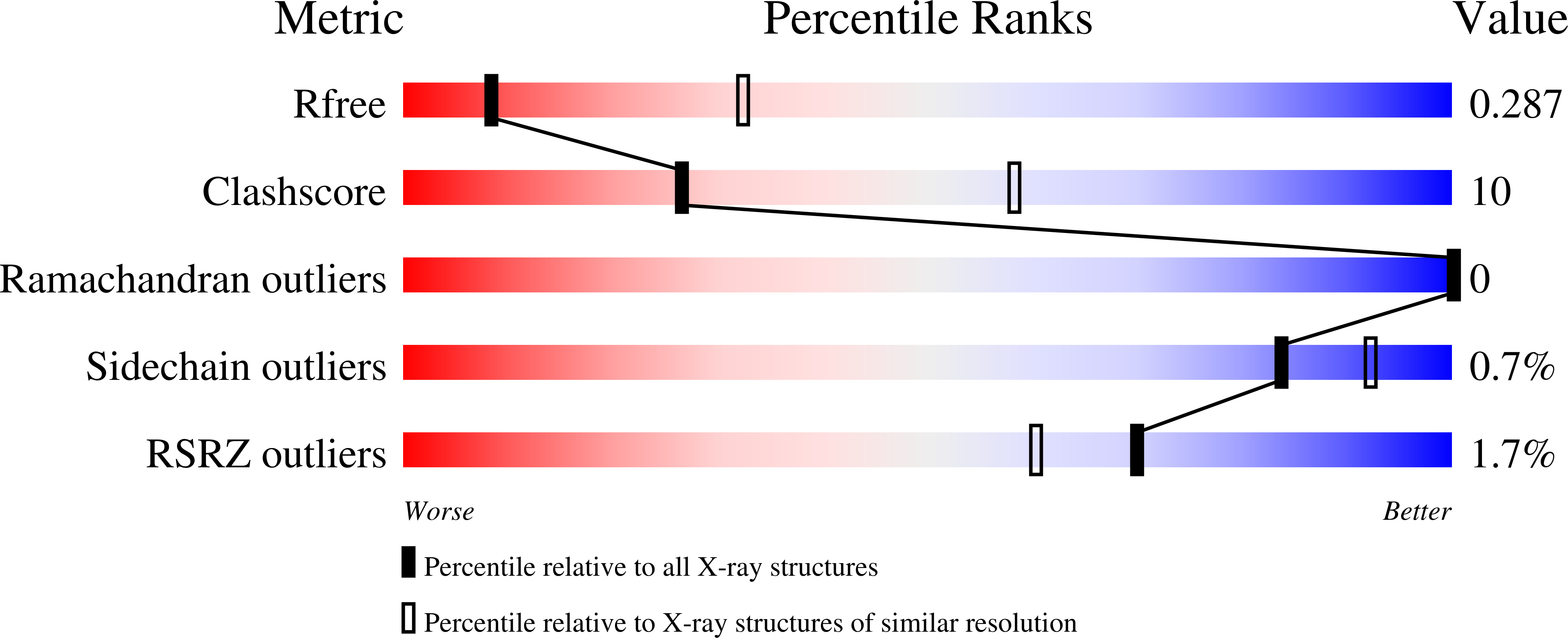
Resolution:
3.23 Å
R-Value Free:
0.28
R-Value Work:
0.23
R-Value Observed:
0.24
Space Group:
P 21 21 21
Macromolecular Entities
Structures with similar UniProt ID
Protein Blast
Polymer Type:polypeptide(L)
Molecule:Receptor-interacting serine/threonine-protein kinase 3
Gene (Uniprot):RIPK3
Mutagens:C3S, C110A
Chain IDs:A, B, C, D
Chain Length:319
Number of Molecules:4
Biological Source:Homo sapiens
Primary Citation
Human RIPK3 maintains MLKL in an inactive conformation prior to cell death by necroptosis.
Nat Commun 12 6783 6783 (2021)
PMID: 34811356 DOI: 10.1038/s41467-021-27032-x

Abstact

The ancestral origins of the lytic cell death mode, necroptosis, lie in host defense. However, the dysregulation of necroptosis in inflammatory diseases has led to widespread interest in targeting the pathway therapeutically. This mode of cell death is executed by the terminal effector, the MLKL pseudokinase, which is licensed to kill following phosphorylation by its upstream regulator, RIPK3 kinase. The precise molecular details underlying MLKL activation are still emerging and, intriguingly, appear to mechanistically-diverge between species. Here, we report the structure of the human RIPK3 kinase domain alone and in complex with the MLKL pseudokinase. These structures reveal how human RIPK3 structurally differs from its mouse counterpart, and how human RIPK3 maintains MLKL in an inactive conformation prior to induction of necroptosis. Residues within the RIPK3:MLKL C-lobe interface are crucial to complex assembly and necroptotic signaling in human cells, thereby rationalizing the strict species specificity governing RIPK3 activation of MLKL.

Legend

Protein

Chemical

Disease

Primary Citation of related structures
Feedback Form
Name
Email
Institute
Feedback