7MT5 image
Entry Detail
PDB ID:
7MT5
Keywords:
Title:
Crystal structure of tryptophan synthase in complex with F9, Cs+, pH7.8 - alpha aminoacrylate form - E(A-A)
Biological Source:
Source Organism:
Host Organism:
PDB Version:
Deposition Date:
2021-05-12
Release Date:
2021-12-29
Method Details:
Experimental Method:
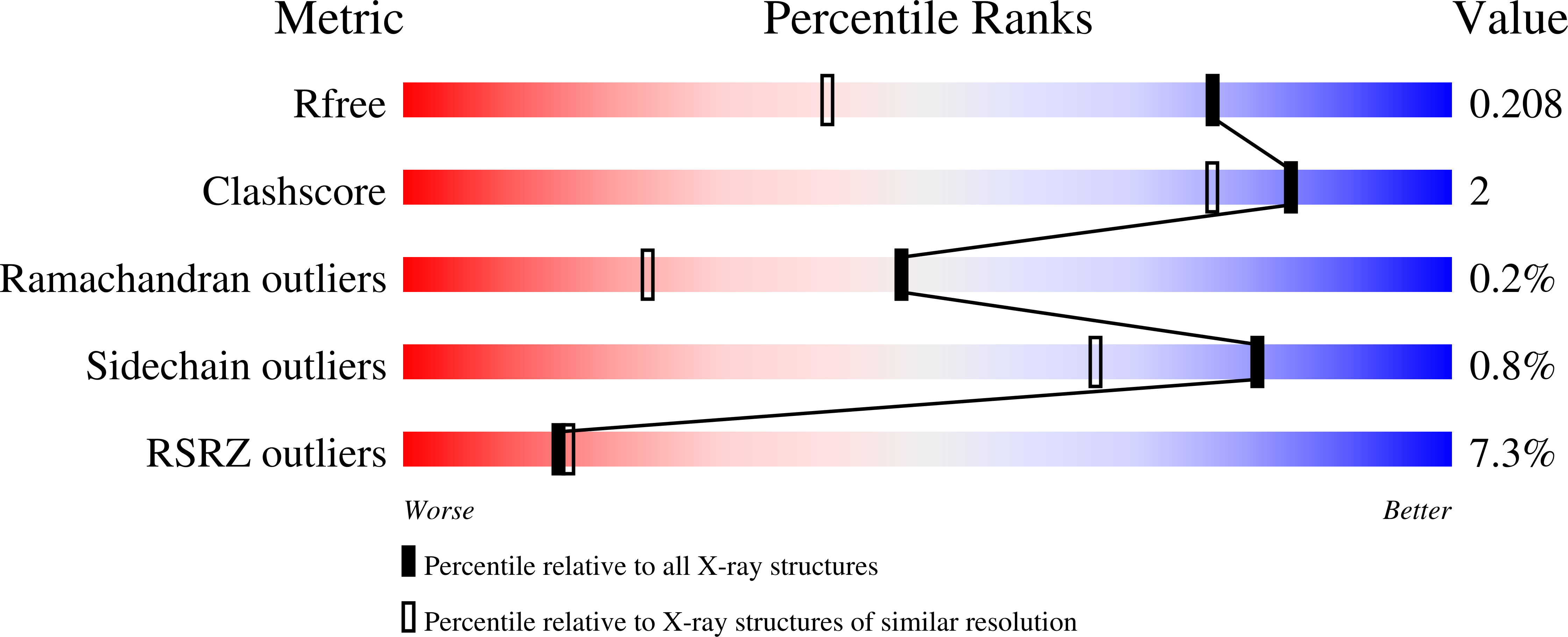
Resolution:
1.50 Å
R-Value Free:
0.20
R-Value Work:
0.17
Space Group:
C 1 2 1
Macromolecular Entities
Polymer Type:polypeptide(L)
Description:Tryptophan synthase alpha chain
Chain IDs:A
Chain Length:268
Number of Molecules:1
Biological Source:Salmonella typhimurium
Polymer Type:polypeptide(L)
Description:Tryptophan synthase beta chain
Chain IDs:B
Chain Length:397
Number of Molecules:1
Biological Source:Salmonella typhimurium
Primary Citation
Imaging active site chemistry and protonation states: NMR crystallography of the tryptophan synthase alpha-aminoacrylate intermediate.
Proc.Natl.Acad.Sci.USA 119 ? ? (2022)
PMID: 34996869 DOI: 10.1073/pnas.2109235119

Abstact

NMR-assisted crystallography-the integrated application of solid-state NMR, X-ray crystallography, and first-principles computational chemistry-holds significant promise for mechanistic enzymology: by providing atomic-resolution characterization of stable intermediates in enzyme active sites, including hydrogen atom locations and tautomeric equilibria, NMR crystallography offers insight into both structure and chemical dynamics. Here, this integrated approach is used to characterize the tryptophan synthase α-aminoacrylate intermediate, a defining species for pyridoxal-5'-phosphate-dependent enzymes that catalyze β-elimination and replacement reactions. For this intermediate, NMR-assisted crystallography is able to identify the protonation states of the ionizable sites on the cofactor, substrate, and catalytic side chains as well as the location and orientation of crystallographic waters within the active site. Most notable is the water molecule immediately adjacent to the substrate β-carbon, which serves as a hydrogen bond donor to the ε-amino group of the acid-base catalytic residue βLys87. From this analysis, a detailed three-dimensional picture of structure and reactivity emerges, highlighting the fate of the L-serine hydroxyl leaving group and the reaction pathway back to the preceding transition state. Reaction of the α-aminoacrylate intermediate with benzimidazole, an isostere of the natural substrate indole, shows benzimidazole bound in the active site and poised for, but unable to initiate, the subsequent bond formation step. When modeled into the benzimidazole position, indole is positioned with C3 in contact with the α-aminoacrylate Cβ and aligned for nucleophilic attack. Here, the chemically detailed, three-dimensional structure from NMR-assisted crystallography is key to understanding why benzimidazole does not react, while indole does.

Legend

Protein

Chemical

Disease

Primary Citation of related structures