7MPA image
Deposition Date 2021-05-04
Release Date 2021-06-30
Last Version Date 2024-05-15
Entry Detail
PDB ID:
7MPA
Title:
Structure and topology of DWORF in bicelles by oriented solid-state NMR
Biological Source:
Source Organism:
Homo sapiens (Taxon ID: 9606)
Host Organism:
Method Details:
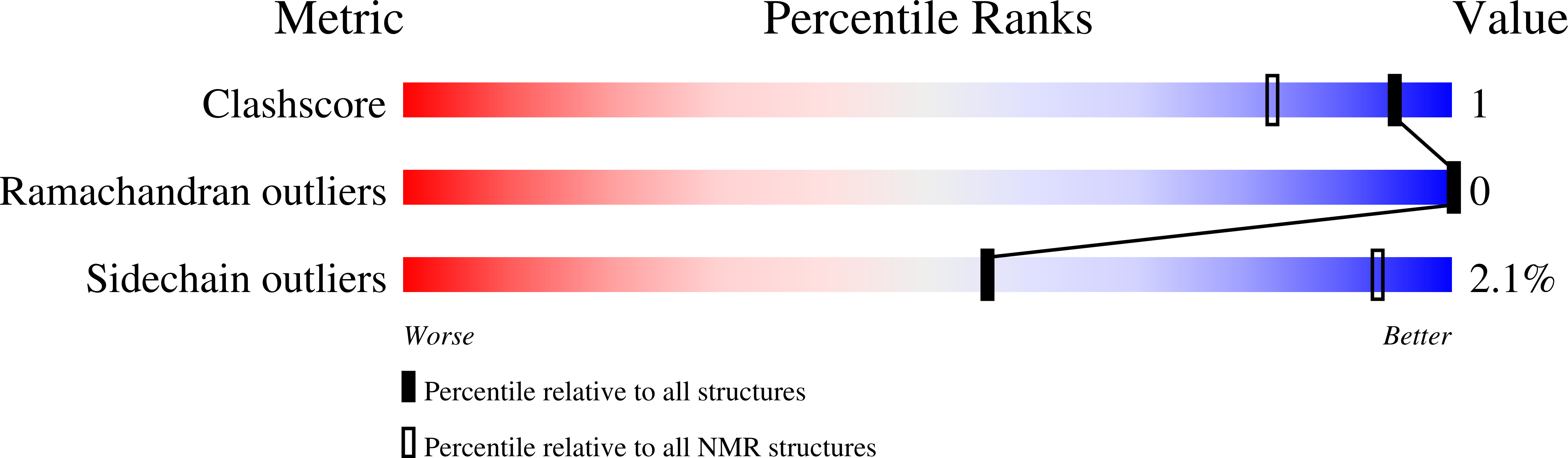
Experimental Method:
Conformers Calculated:
5000
Conformers Submitted:
10
Selection Criteria:
structures with the lowest energy
Macromolecular Entities
Structures with similar UniProt ID
Protein Blast
Polymer Type:polypeptide(L)
Molecule:Sarcoplasmic/endoplasmic reticulum calcium ATPase regulator DWORF
Gene (Uniprot):STRIT1
Chain IDs:A
Chain Length:36
Number of Molecules:1
Biological Source:Homo sapiens
Ligand Molecules
Primary Citation
A kink in DWORF helical structure controls the activation of the sarcoplasmic reticulum Ca 2+ -ATPase.
Structure 30 360 ? (2022)
PMID: 34875216 DOI: 10.1016/j.str.2021.11.003

Abstact

SERCA is a P-type ATPase embedded in the sarcoplasmic reticulum and plays a central role in muscle relaxation. SERCA's function is regulated by single-pass membrane proteins called regulins. Unlike other regulins, dwarf open reading frame (DWORF) expressed in cardiac muscle has a unique activating effect. Here, we determine the structure and topology of DWORF in lipid bilayers using a combination of oriented sample solid-state NMR spectroscopy and replica-averaged orientationally restrained molecular dynamics. We found that DWORF's structural topology consists of a dynamic N-terminal domain, an amphipathic juxtamembrane helix that crosses the lipid groups at an angle of 64°, and a transmembrane C-terminal helix with an angle of 32°. A kink induced by Pro15, unique to DWORF, separates the two helical domains. A single Pro15Ala mutant significantly decreases the kink and eliminates DWORF's activating effect on SERCA. Overall, our findings directly link DWORF's structural topology to its activating effect on SERCA.

Legend

Protein

Chemical

Disease

Primary Citation of related structures