7MP8 image
Entry Detail
PDB ID:
7MP8
Title:
Crystal structure of the cytosolic domain of Tribolium castaneum PINK1 in the non-phosphorylated state
Biological Source:
Source Organism:
Host Organism:
PDB Version:
Deposition Date:
2021-05-04
Release Date:
2021-12-01
Method Details:
Experimental Method:
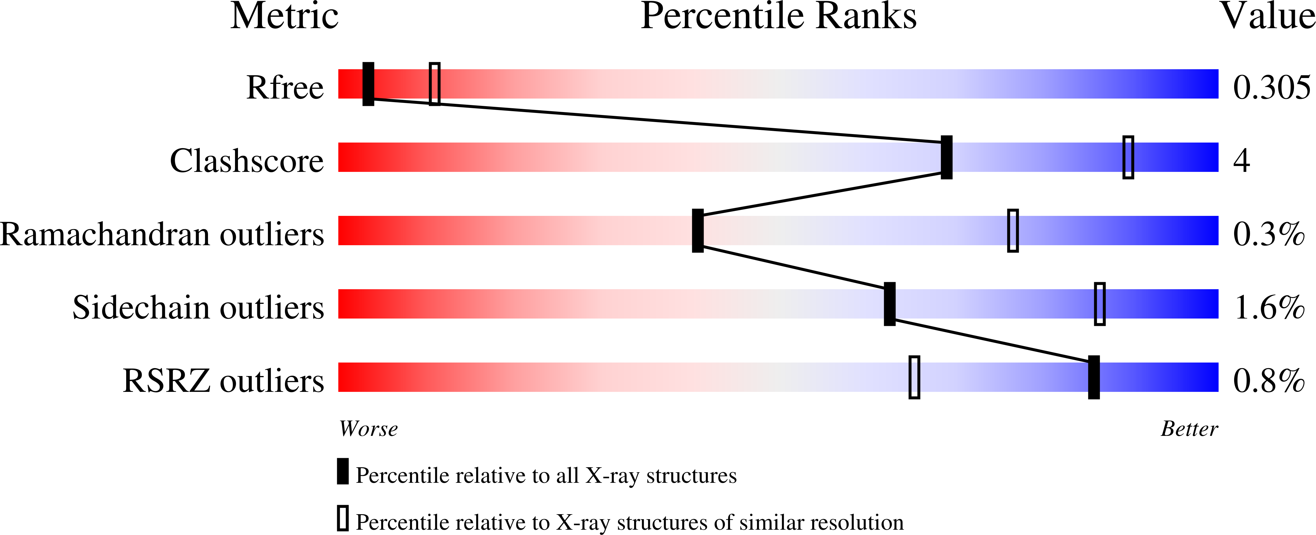
Resolution:
3.00 Å
R-Value Free:
0.30
R-Value Work:
0.25
R-Value Observed:
0.25
Space Group:
P 61 2 2
Macromolecular Entities
Polymer Type:polypeptide(L)
Description:Serine/threonine-protein kinase PINK1, mitochondrial-like Protein
Chain IDs:A
Chain Length:455
Number of Molecules:1
Biological Source:Tribolium castaneum
Ligand Molecules
Primary Citation
Mechanism of PINK1 activation by autophosphorylation and insights into assembly on the TOM complex.
Mol.Cell 82 44 ? (2022)
PMID: 34875213 DOI: 10.1016/j.molcel.2021.11.012

Abstact

Mutations in PINK1 cause autosomal-recessive Parkinson's disease. Mitochondrial damage results in PINK1 import arrest on the translocase of the outer mitochondrial membrane (TOM) complex, resulting in the activation of its ubiquitin kinase activity by autophosphorylation and initiation of Parkin-dependent mitochondrial clearance. Herein, we report crystal structures of the entire cytosolic domain of insect PINK1. Our structures reveal a dimeric autophosphorylation complex targeting phosphorylation at the invariant Ser205 (human Ser228). The dimer interface requires insert 2, which is unique to PINK1. The structures also reveal how an N-terminal helix binds to the C-terminal extension and provide insights into stabilization of PINK1 on the core TOM complex.

Legend

Protein

Chemical

Disease

Primary Citation of related structures