7MLU image
Deposition Date 2021-04-29
Release Date 2021-09-29
Last Version Date 2024-11-13
Entry Detail
PDB ID:
7MLU
Title:
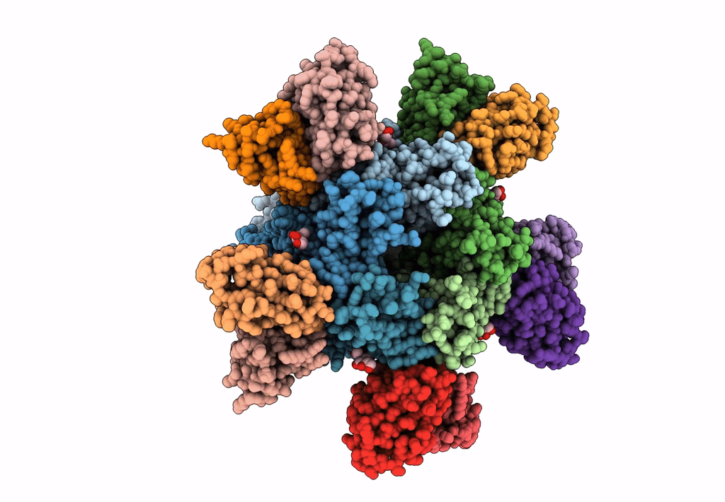
Cryo-EM reveals partially and fully assembled native glycine receptors,homomeric pentamer
Biological Source:
Source Organism:
Rattus norvegicus (Taxon ID: 10116)
Sus scrofa (Taxon ID: 9823)
Host Organism:
Method Details:
Experimental Method:
Resolution:
4.10 Å
Aggregation State:
PARTICLE
Reconstruction Method:
SINGLE PARTICLE
Macromolecular Entities
Structures with similar UniProt ID
Protein Blast
Polymer Type:polypeptide(L)
Molecule:Glycine receptor alpha 1
Chain IDs:K (auth: A), L (auth: E), M (auth: D), N (auth: B), O (auth: C)
Chain Length:456
Number of Molecules:5
Biological Source:Sus scrofa
Polymer Type:polypeptide(L)
Molecule:3D1 Fab Heavy Chain
Chain IDs:B (auth: F), D (auth: G), F (auth: N), H (auth: L), J (auth: H)
Chain Length:118
Number of Molecules:5
Biological Source:Rattus norvegicus
Polymer Type:polypeptide(L)
Molecule:3D1 Fab Light Chain
Chain IDs:A (auth: K), C (auth: J), E (auth: O), G (auth: M), I
Chain Length:107
Number of Molecules:5
Biological Source:Rattus norvegicus
Ligand Molecules
Primary Citation
Architecture and assembly mechanism of native glycine receptors.
Nature 599 513 517 (2021)
PMID: 34555840 DOI: 10.1038/s41586-021-04022-z

Abstact

Glycine receptors (GlyRs) are pentameric, 'Cys-loop' receptors that form chloride-permeable channels and mediate fast inhibitory signalling throughout the central nervous system1,2. In the spinal cord and brainstem, GlyRs regulate locomotion and cause movement disorders when mutated2,3. However, the stoichiometry of native GlyRs and the mechanism by which they are assembled remain unclear, despite extensive investigation4-8. Here we report cryo-electron microscopy structures of native GlyRs from pig spinal cord and brainstem, revealing structural insights into heteromeric receptors and their predominant subunit stoichiometry of 4α:1β. Within the heteromeric pentamer, the β(+)-α(-) interface adopts a structure that is distinct from the α(+)-α(-) and α(+)-β(-) interfaces. Furthermore, the β-subunit contains a unique phenylalanine residue that resides within the pore and disrupts the canonical picrotoxin site. These results explain why inclusion of the β-subunit breaks receptor symmetry and alters ion channel pharmacology. We also find incomplete receptor complexes and, by elucidating their structures, reveal the architectures of partially assembled α-trimers and α-tetramers.

Legend

Protein

Chemical

Disease

Primary Citation of related structures