
Deposition Date
2021-04-18
Release Date
2021-07-07
Last Version Date
2025-05-21
Entry Detail
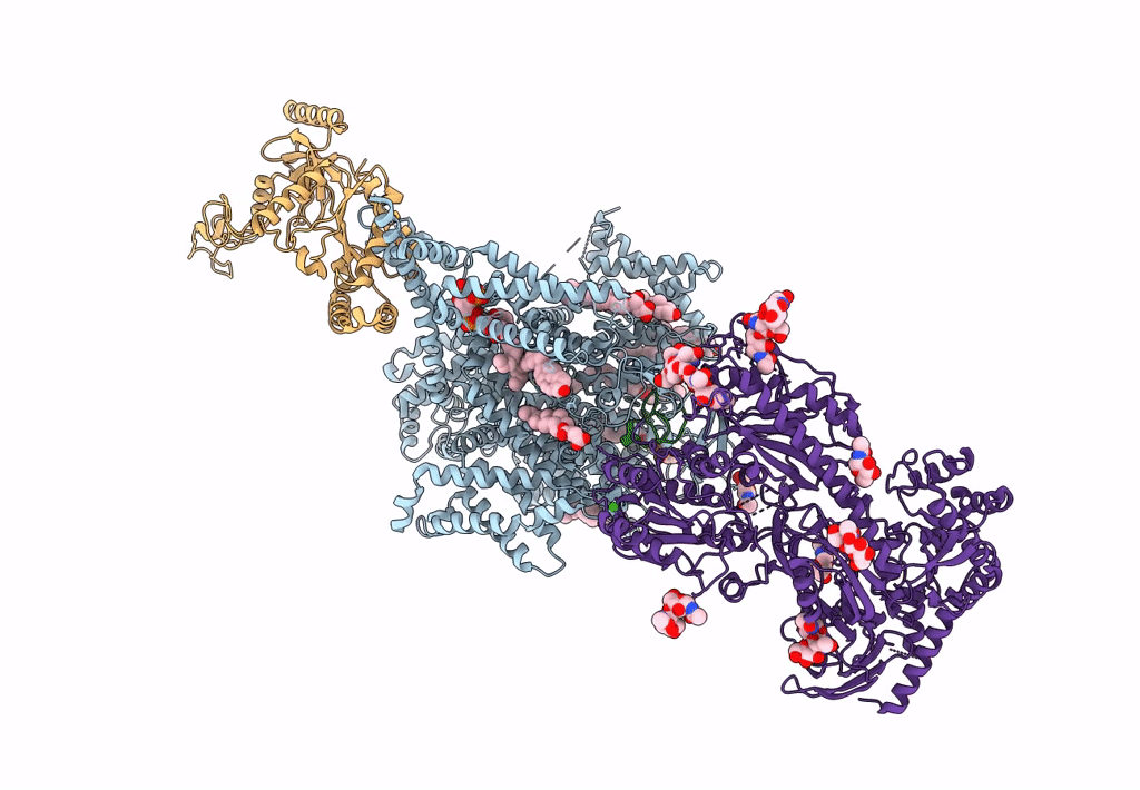
PDB ID:
7MIX
Keywords:
Title:
Human N-type voltage-gated calcium channel Cav2.2 in the presence of ziconotide at 3.0 Angstrom resolution
Biological Source:
Source Organism(s):
Homo sapiens (Taxon ID: 9606)
Conus magus (Taxon ID: 6492)
Conus magus (Taxon ID: 6492)
Expression System(s):
Method Details:
Experimental Method:
Resolution:
3.00 Å
Aggregation State:
PARTICLE
Reconstruction Method:
SINGLE PARTICLE
