
Deposition Date
2021-04-14
Release Date
2021-12-29
Last Version Date
2023-10-18
Entry Detail
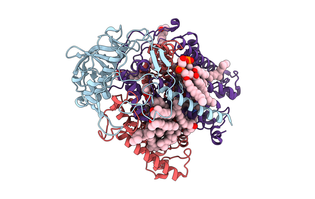
PDB ID:
7MH3
Keywords:
Title:
Crystal structure of R. sphaeroides Photosynthetic Reaction Center variant; Y(M210)3-chlorotyrosine
Biological Source:
Source Organism(s):
Rhodobacter sphaeroides (Taxon ID: 1063)
Expression System(s):
Method Details:
Experimental Method:
Resolution:
2.30 Å
R-Value Free:
0.17
R-Value Work:
0.15
R-Value Observed:
0.15
Space Group:
P 31 2 1
