
Deposition Date
2021-04-08
Release Date
2022-01-05
Last Version Date
2024-05-29
Entry Detail
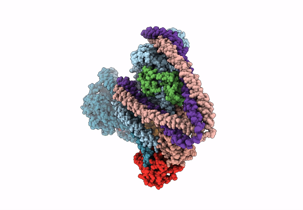
PDB ID:
7MF3
Keywords:
Title:
Structure of the autoinhibited state of smooth muscle myosin-2
Biological Source:
Source Organism(s):
Gallus gallus (Taxon ID: 9031)
Method Details:
Experimental Method:
Resolution:
3.40 Å
Aggregation State:
TISSUE
Reconstruction Method:
SINGLE PARTICLE
