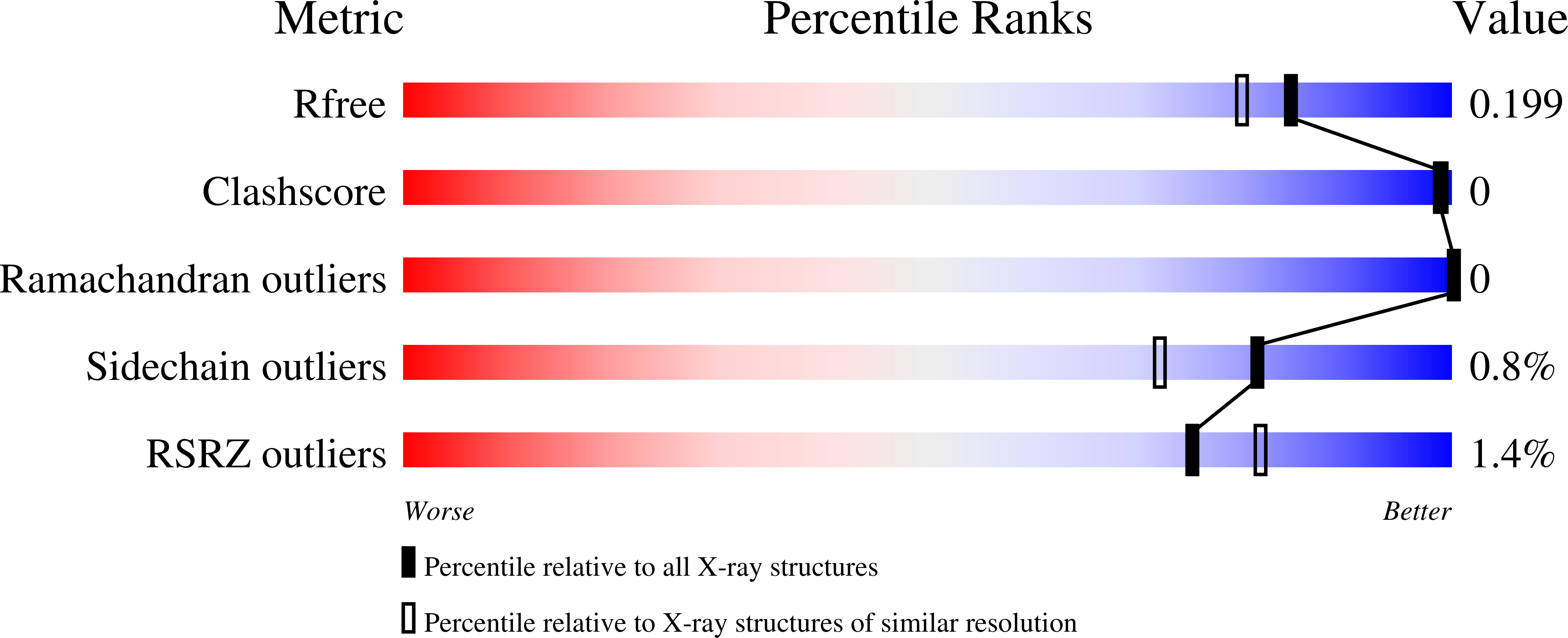
7MEL image
Deposition Date 2021-04-06
Release Date 2021-07-14
Last Version Date 2023-10-18
Entry Detail
PDB ID:
7MEL
Keywords:
Title:
Sco GlgEI-V279S in complex with 4-alpha-glucoside of validamine
Biological Source:
Source Organism:
Host Organism:
Method Details:
Experimental Method:
Resolution:
1.75 Å
R-Value Free:
0.19
R-Value Work:
0.17
R-Value Observed:
0.17
Space Group:
P 41 21 2
Macromolecular Entities
Polymer Type:polypeptide(L)
Molecule:Alpha-1,4-glucan:maltose-1-phosphate maltosyltransferase 1
Gene (Uniprot):glgE1
Chain IDs:A, B
Chain Length:683
Number of Molecules:2
Biological Source:Streptomyces coelicolor
Primary Citation
Stereoselective synthesis of a 4-alpha-glucoside of valienamine and its X-ray structure in complex with Streptomyces coelicolor GlgE1-V279S.
Sci Rep 11 13413 13413 (2021)
PMID: 34183716 DOI: 10.1038/s41598-021-92554-9

Abstact

Glycoside hydrolases (GH) are a large family of hydrolytic enzymes found in all domains of life. As such, they control a plethora of normal and pathogenic biological functions. Thus, understanding selective inhibition of GH enzymes at the atomic level can lead to the identification of new classes of therapeutics. In these studies, we identified a 4-⍺-glucoside of valienamine (8) as an inhibitor of Streptomyces coelicolor (Sco) GlgE1-V279S which belongs to the GH13 Carbohydrate Active EnZyme family. The results obtained from the dose-response experiments show that 8 at a concentration of 1000 µM reduced the enzyme activity of Sco GlgE1-V279S by 65%. The synthetic route to 8 and a closely related 4-⍺-glucoside of validamine (7) was achieved starting from readily available D-maltose. A key step in the synthesis was a chelation-controlled addition of vinylmagnesium bromide to a maltose-derived enone intermediate. X-ray structures of both 7 and 8 in complex with Sco GlgE1-V279S were solved to resolutions of 1.75 and 1.83 Å, respectively. Structural analysis revealed the valienamine derivative 8 binds the enzyme in an E2 conformation for the cyclohexene fragment. Also, the cyclohexene fragment shows a new hydrogen-bonding contact from the pseudo-diaxial C(3)-OH to the catalytic nucleophile Asp 394 at the enzyme active site. Asp 394, in fact, forms a bidentate interaction with both the C(3)-OH and C(7)-OH of the inhibitor. In contrast, compound 7 disrupts the catalytic sidechain interaction network of Sco GlgE1-V279S via steric interactions resulting in a conformation change in Asp 394. These findings will have implications for the design other aminocarbasugar-based GH13-inhibitors and will be useful for identifying more potent and selective inhibitors.

Legend

Protein

Chemical

Disease

Primary Citation of related structures