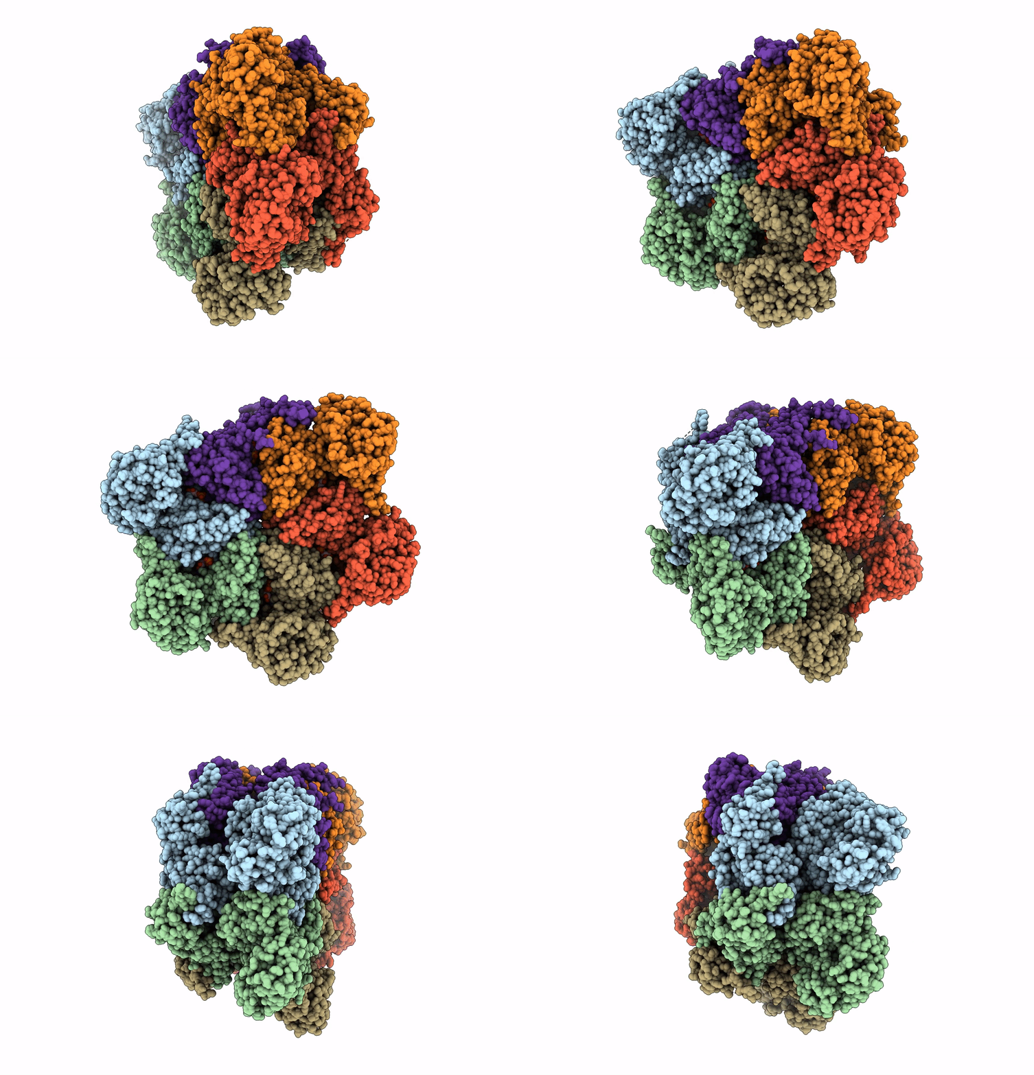
7MDM image
Entry Detail
PDB ID:
7MDM
EMDB ID:
Keywords:
Title:
Structure of human p97 ATPase L464P mutant
Biological Source:
Source Organism:
Host Organism:
PDB Version:
Deposition Date:
2021-04-05
Release Date:
2021-08-25
Method Details:
Experimental Method:
Resolution:
4.86 Å
Aggregation State:
PARTICLE
Reconstruction Method:
SINGLE PARTICLE
Macromolecular Entities
Polymer Type:polypeptide(L)
Description:Transitional endoplasmic reticulum ATPase
Chain IDs:A (auth: B), B (auth: C), C (auth: D), D (auth: E), E (auth: A), F
Chain Length:806
Number of Molecules:6
Biological Source:Homo sapiens
Ligand Molecules
Primary Citation
Conserved L464 in p97 D1-D2 linker is critical for p97 cofactor regulated ATPase activity.
Biochem.J. 478 3185 3204 (2021)
PMID: 34405853 DOI: 10.1042/BCJ20210288

Abstact

p97 protein is a highly conserved, abundant, functionally diverse, structurally dynamic homohexameric AAA enzyme-containing N, D1, and D2 domains. A truncated p97 protein containing the N and D1 domains and the D1-D2 linker (ND1L) exhibits 79% of wild-type (WT) ATPase activity whereas the ND1 domain alone without the linker only has 2% of WT activity. To investigate the relationship between the D1-D2 linker and the D1 domain, we produced p97 ND1L mutants and demonstrated that this 22-residue linker region is essential for D1 ATPase activity. The conserved amino acid leucine 464 (L464) is critical for regulating D1 and D2 ATPase activity by p97 cofactors p37, p47, and Npl4-Ufd1 (NU). Changing leucine to alanine, proline, or glutamate increased the maximum rate of ATP turnover (kcat) of p47-regulated ATPase activities for these mutants, but not for WT. p37 and p47 increased the kcat of the proline substituted linker, suggesting that they induced linker conformations facilitating ATP hydrolysis. NU inhibited D1 ATPase activities of WT and mutant ND1L proteins, but activated D2 ATPase activity of full-length p97. To further understand the mutant mechanism, we used single-particle cryo-EM to visualize the full-length p97L464P and revealed the conformational change of the D1-D2 linker, resulting in a movement of the helix-turn-helix motif (543-569). Taken together with the biochemical and structural results we conclude that the linker helps maintain D1 in a competent conformation and relays the communication to/from the N-domain to the D1 and D2 ATPase domains, which are ∼50 Å away.

Legend

Protein

Chemical

Disease

Primary Citation of related structures