Deposition Date
2021-03-27
Release Date
2021-09-01
Last Version Date
2024-10-30
Entry Detail
PDB ID:
7M7D
Keywords:
Title:
Crystal structure of the indoleamine 2,3-dioxygenagse 1 (IDO1) complexed with IACS-8968
Biological Source:
Source Organism(s):
Homo sapiens (Taxon ID: 9606)
Expression System(s):
Method Details:
Experimental Method:
Resolution:
2.60 Å
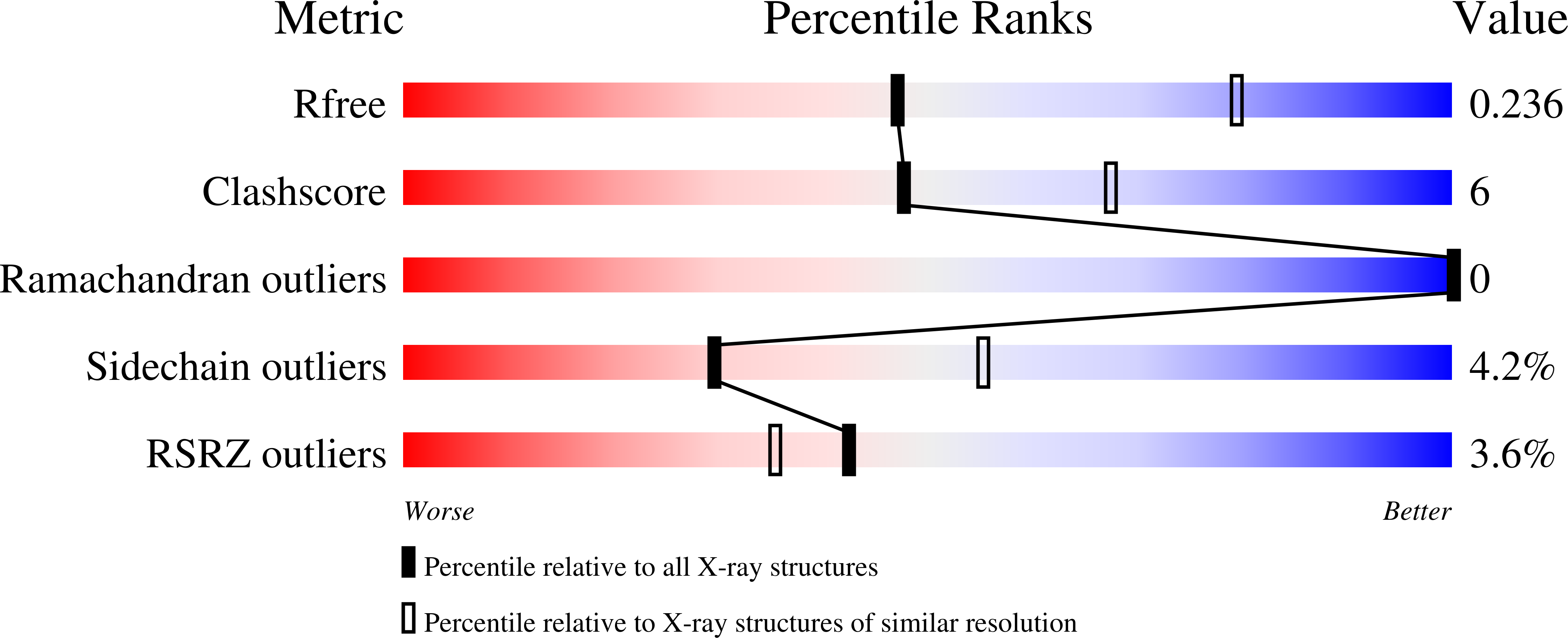
R-Value Free:
0.23
R-Value Work:
0.20
R-Value Observed:
0.21
Space Group:
P 21 21 21