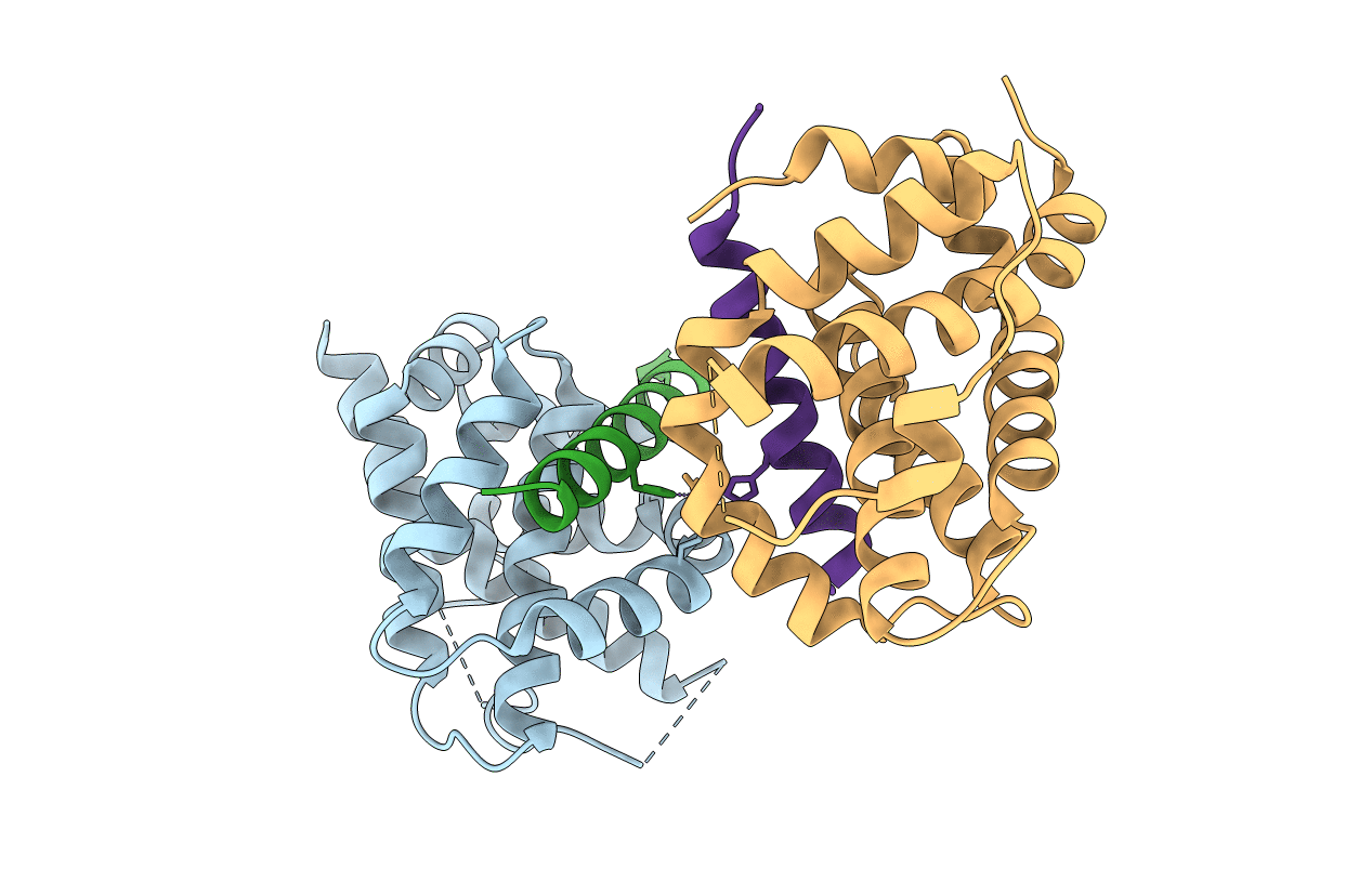
7M5B image
Entry Detail
PDB ID:
7M5B
Keywords:
Title:
Crystal Structure of human BAK in complex with M3W5_BID
Biological Source:
Source Organism:
Host Organism:
PDB Version:
Deposition Date:
2021-03-23
Release Date:
2022-01-12
Method Details:
Experimental Method:
Resolution:
1.85 Å
R-Value Free:
0.20
R-Value Work:
0.16
R-Value Observed:
0.17
Space Group:
P 21 21 21
Macromolecular Entities
Polymer Type:polypeptide(L)
Description:Bcl-2 homologous antagonist/killer
Chain IDs:A, C
Chain Length:166
Number of Molecules:2
Biological Source:Homo sapiens
Polymer Type:polypeptide(L)
Description:BH3-interacting domain death agonist p15
Chain IDs:B, D
Chain Length:25
Number of Molecules:2
Biological Source:Homo sapiens
Ligand Molecules
Primary Citation
Structural basis of BAK activation in mitochondrial apoptosis initiation.
Nat Commun 13 250 250 (2022)
PMID: 35017502 DOI: 10.1038/s41467-021-27851-y

Abstact

BCL-2 proteins regulate mitochondrial poration in apoptosis initiation. How the pore-forming BCL-2 Effector BAK is activated remains incompletely understood mechanistically. Here we investigate autoactivation and direct activation by BH3-only proteins, which cooperate to lower BAK threshold in membrane poration and apoptosis initiation. We define in trans BAK autoactivation as the asymmetric "BH3-in-groove" triggering of dormant BAK by active BAK. BAK autoactivation is mechanistically similar to direct activation. The structure of autoactivated BAK BH3-BAK complex reveals the conformational changes leading to helix α1 destabilization, which is a hallmark of BAK activation. Helix α1 is destabilized and restabilized in structures of BAK engaged by rationally designed, high-affinity activating and inactivating BID-like BH3 ligands, respectively. Altogether our data support the long-standing hit-and-run mechanism of BAK activation by transient binding of BH3-only proteins, demonstrating that BH3-induced structural changes are more important in BAK activation than BH3 ligand affinity.

Legend

Protein

Chemical

Disease

Primary Citation of related structures