Abstact
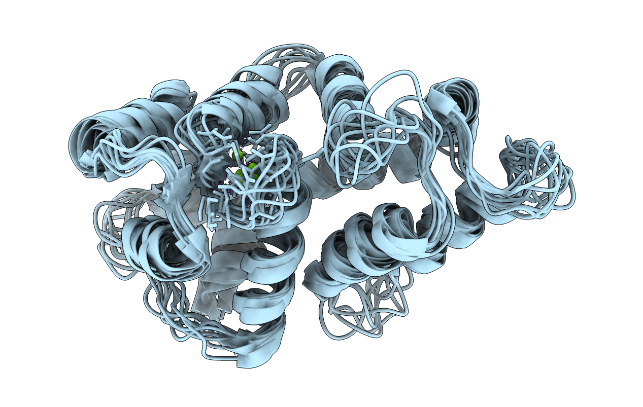
Retinal guanylate cyclases (RetGCs) are regulated by a family of guanylate cyclase-activating proteins (called GCAP1-7). GCAPs form dimers that bind to Ca2+ and confer Ca2+ sensitive activation of RetGC during visual phototransduction. The GCAP5 homologue from zebrafish contains two nonconserved cysteine residues (Cys15 and Cys17) that bind to ferrous ion, which stabilizes GCAP5 dimerization and diminishes its ability to activate RetGC. Here, we present NMR and EPR-DEER structural analysis of a GCAP5 dimer in the Mg2+-bound, Ca2+-free, Fe2+-free activator state. The NMR-derived structure of GCAP5 is similar to the crystal structure of Ca2+-bound GCAP1 (root-mean-square deviation of 2.4 Å), except that the N-terminal helix of GCAP5 is extended by two residues, which allows the sulfhydryl groups of Cys15 and Cys17 to become more solvent exposed in GCAP5 to facilitate Fe2+ binding. Nitroxide spin-label probes were covalently attached to particular cysteine residues engineered in GCAP5: C15, C17, T26C, C28, N56C, C69, C105, N139C, E152C, and S159C. The intermolecular distance of each spin-label probe in dimeric GCAP5 (measured by EPR-DEER) defined restraints for calculating the dimer structure by molecular docking. The GCAP5 dimer possesses intermolecular hydrophobic contacts involving the side chain atoms of H18, Y21, M25, F72, V76, and W93, as well as an intermolecular salt bridge between R22 and D71. The structural model of the GCAP5 dimer was validated by mutations (H18E/Y21E, H18A/Y21A, R22D, R22A, M25E, D71R, F72E, and V76E) at the dimer interface that disrupt dimerization of GCAP5 and affect the activation of RetGC. We propose that GCAP5 dimerization may play a role in the Fe2+-dependent regulation of cyclase activity in zebrafish photoreceptors.


