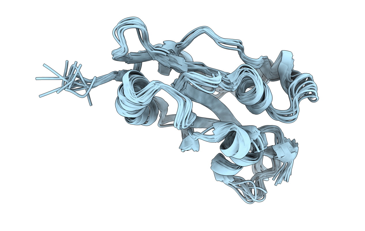
7M0G image
Entry Detail
PDB ID:
7M0G
Title:
Magic Angle Spinning NMR Structure of Human Cofilin-2 Assembled on Actin Filaments
Biological Source:
Source Organism:
Host Organism:
PDB Version:
Deposition Date:
2021-03-10
Release Date:
2022-04-20
Method Details:
Experimental Method:
Conformers Calculated:
250
Conformers Submitted:
10
Selection Criteria:
structures with the lowest energy
Macromolecular Entities
Polymer Type:polypeptide(L)
Description:Cofilin-2
Chain IDs:A
Chain Length:166
Number of Molecules:1
Biological Source:Homo sapiens
Ligand Molecules
Primary Citation
Magic angle spinning NMR structure of human cofilin-2 assembled on actin filaments reveals isoform-specific conformation and binding mode.
Nat Commun 13 2114 2114 (2022)
PMID: 35440100 DOI: 10.1038/s41467-022-29595-9

Abstact

Actin polymerization dynamics regulated by actin-binding proteins are essential for various cellular functions. The cofilin family of proteins are potent regulators of actin severing and filament disassembly. The structural basis for cofilin-isoform-specific severing activity is poorly understood as their high-resolution structures in complex with filamentous actin (F-actin) are lacking. Here, we present the atomic-resolution structure of the muscle-tissue-specific isoform, cofilin-2 (CFL2), assembled on ADP-F-actin, determined by magic-angle-spinning (MAS) NMR spectroscopy and data-guided molecular dynamics (MD) simulations. We observe an isoform-specific conformation for CFL2. This conformation is the result of a unique network of hydrogen bonding interactions within the α2 helix containing the non-conserved residue, Q26. Our results indicate F-site interactions that are specific between CFL2 and ADP-F-actin, revealing mechanistic insights into isoform-dependent F-actin disassembly.

Legend

Protein

Chemical

Disease

Primary Citation of related structures