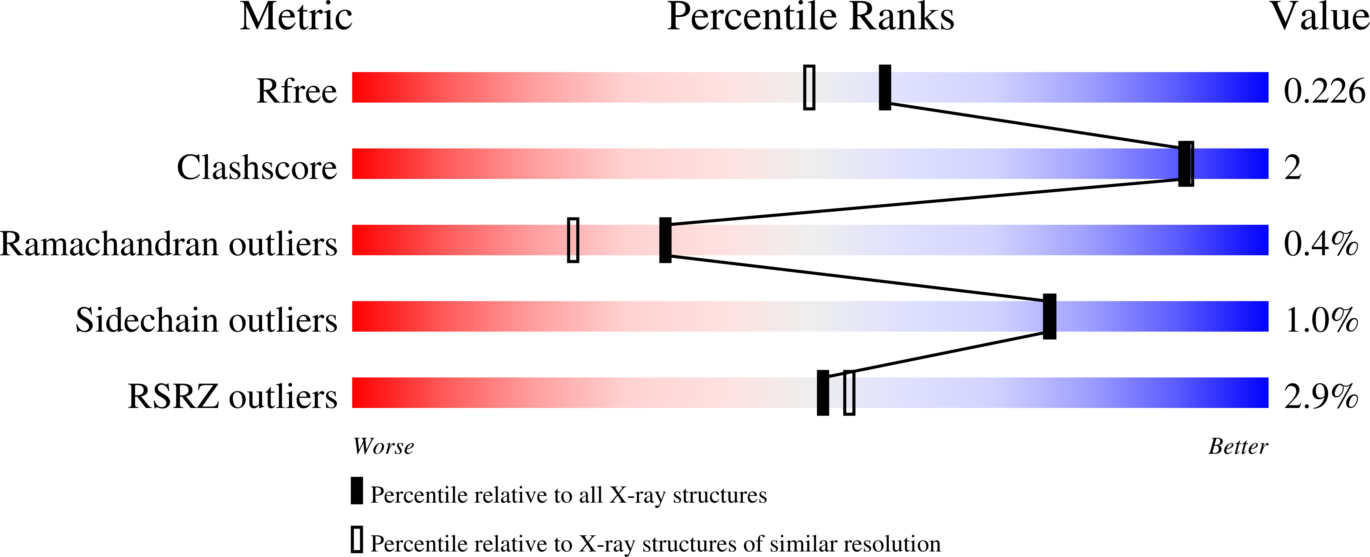
7LVZ image
Deposition Date 2021-02-26
Release Date 2022-01-05
Last Version Date 2024-04-03
Entry Detail
PDB ID:
7LVZ
Keywords:
Title:
Crystal structure of ADO
Biological Source:
Source Organism:
Mus musculus (Taxon ID: 10090)
Host Organism:
Method Details:
Experimental Method:
Resolution:
1.89 Å
R-Value Free:
0.22
R-Value Work:
0.19
R-Value Observed:
0.19
Space Group:
P 21 21 21
Macromolecular Entities
Structures with similar UniProt ID
Protein Blast
Polymer Type:polypeptide(L)
Molecule:2-aminoethanethiol dioxygenase
Gene (Uniprot):Ado
Chain IDs:A, B, C, D
Chain Length:256
Number of Molecules:4
Biological Source:Mus musculus
Primary Citation
The Crystal Structure of Cysteamine Dioxygenase Reveals the Origin of the Large Substrate Scope of This Vital Mammalian Enzyme.
Biochemistry 60 3728 3737 (2021)
PMID: 34762398 DOI: 10.1021/acs.biochem.1c00463

Abstact

We report the crystal structure of the mammalian non-heme iron enzyme cysteamine dioxygenase (ADO) at 1.9 Å resolution, which shows an Fe and three-histidine (3-His) active site situated at the end of a wide substrate access channel. The open approach to the active site is consistent with the recent discovery that ADO catalyzes not only the conversion of cysteamine to hypotaurine but also the oxidation of N-terminal cysteine (Nt-Cys) peptides to their corresponding sulfinic acids as part of the eukaryotic N-degron pathway. Whole-protein models of ADO in complex with either cysteamine or an Nt-Cys peptide, generated using molecular dynamics and quantum mechanics/molecular mechanics calculations, suggest occlusion of access to the active site by peptide substrate binding. This finding highlights the importance of a small tunnel that leads from the opposite face of the enzyme into the active site, providing a path through which co-substrate O2 could access the Fe center. Intriguingly, the entrance to this tunnel is guarded by two Cys residues that may form a disulfide bond to regulate O2 delivery in response to changes in the intracellular redox potential. Notably, the Cys and tyrosine residues shown to be capable of forming a cross-link in human ADO reside ∼7 Å from the iron center. As such, cross-link formation may not be structurally or functionally significant in ADO.

Legend

Protein

Chemical

Disease

Primary Citation of related structures