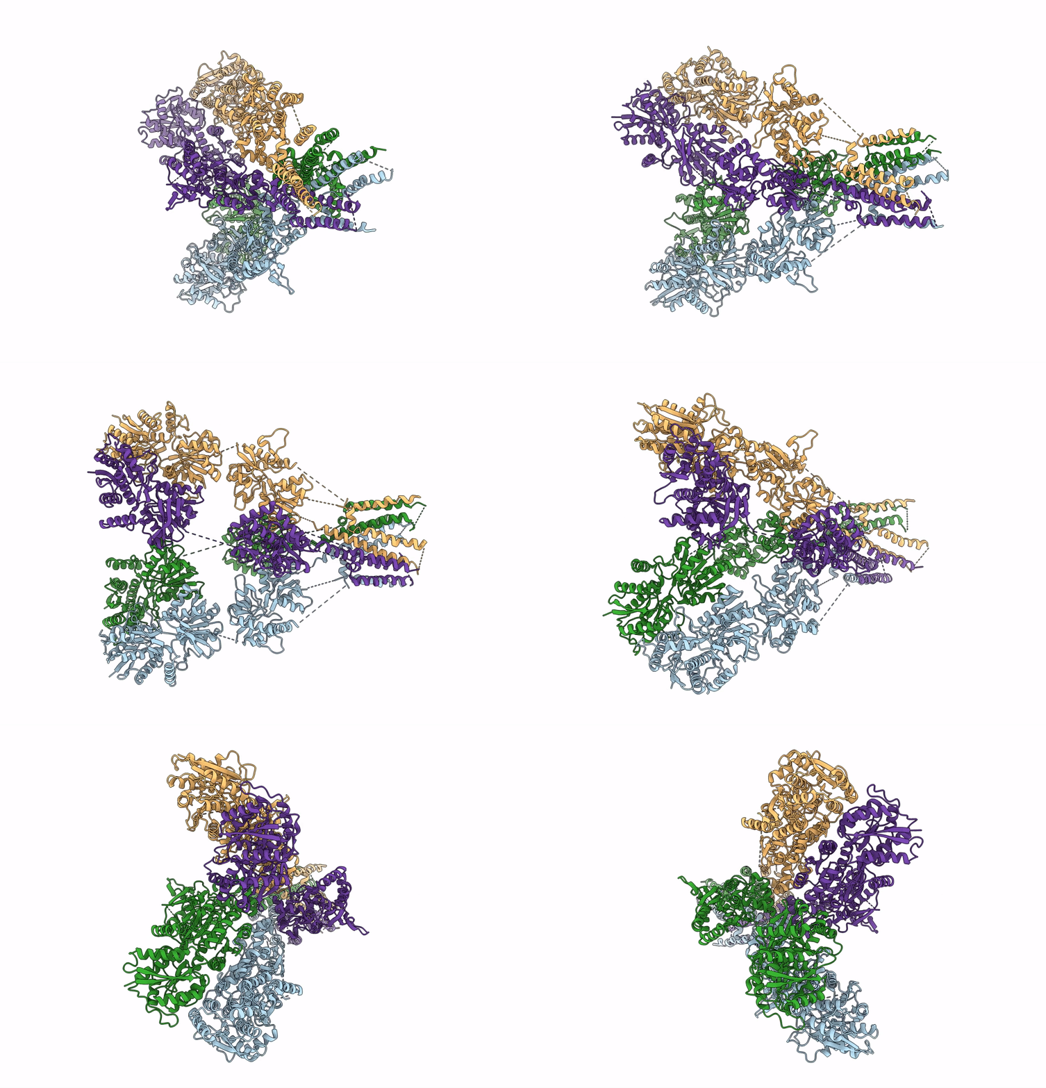
7LVT image
Deposition Date 2021-02-26
Release Date 2021-11-03
Last Version Date 2024-10-09
Entry Detail
PDB ID:
7LVT
Title:
Structure of full-length GluK1 with L-Glu
Biological Source:
Source Organism:
Host Organism:
Method Details:
Experimental Method:
Resolution:
4.60 Å
Aggregation State:
PARTICLE
Reconstruction Method:
SINGLE PARTICLE
Macromolecular Entities
Polymer Type:polypeptide(L)
Molecule:Isoform Glur5-2 of Glutamate receptor ionotropic, kainate 1
Chain IDs:A, B, C, D
Chain Length:904
Number of Molecules:4
Biological Source:Rattus norvegicus
Ligand Molecules
Primary Citation
Structural and compositional diversity in the kainate receptor family.
Cell Rep 37 109891 109891 (2021)
PMID: 34706237 DOI: 10.1016/j.celrep.2021.109891

Abstact

The kainate receptors (KARs) are members of the ionotropic glutamate receptor family and assemble into tetramers from a pool of five subunit types (GluK1-5). Each subunit confers distinct functional properties to a receptor, but the compositional and stoichiometric diversity of KAR tetramers is not well understood. To address this, we first solve the structure of the GluK1 homomer, which enables a systematic assessment of structural compatibility among KAR subunits. Next, we analyze single-cell RNA sequencing data, which reveal extreme diversity in the combinations of two or more KAR subunits co-expressed within the same cell. We then investigate the composition of individual receptor complexes using single-molecule fluorescence techniques and find that di-heteromers assembled from GluK1, GluK2, or GluK3 can form with all possible stoichiometries, while GluK1/K5, GluK2/K5, and GluK3/K5 can form 3:1 or 2:2 complexes. Finally, using three-color single-molecule imaging, we discover that KARs can form tri- and tetra-heteromers.

Legend

Protein

Chemical

Disease

Primary Citation of related structures