Deposition Date
2021-02-05
Release Date
2021-05-12
Last Version Date
2025-05-28
Entry Detail
PDB ID:
7LMB
Keywords:
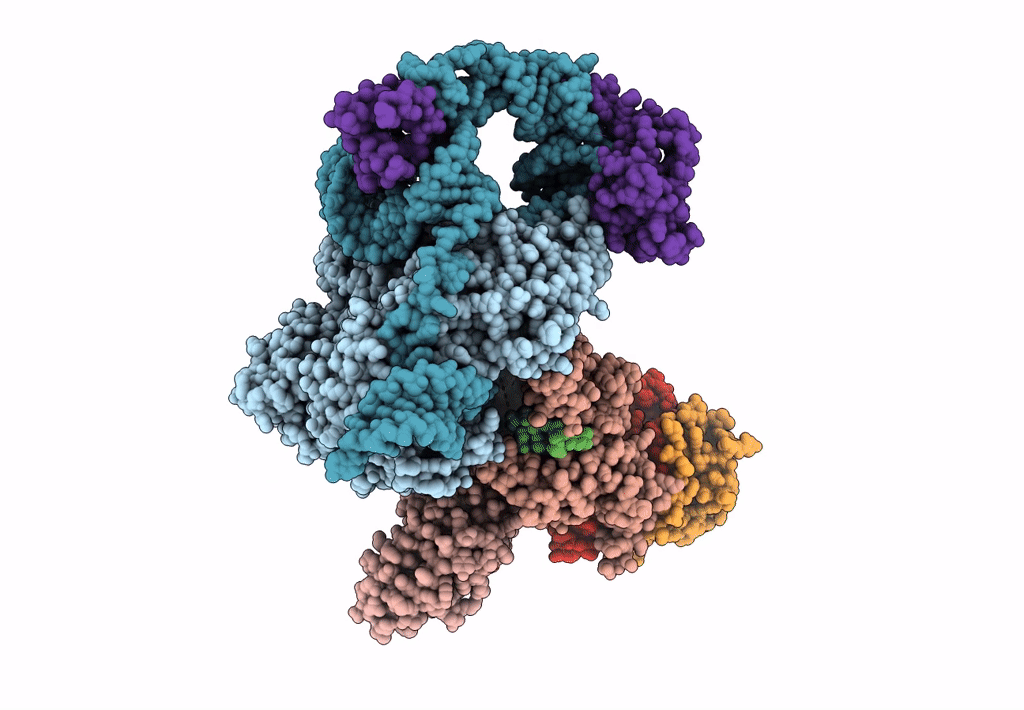
Title:
Tetrahymena telomerase T5D5 structure at 3.8 Angstrom
Biological Source:
Source Organism(s):
Tetrahymena thermophila (Taxon ID: 5911)
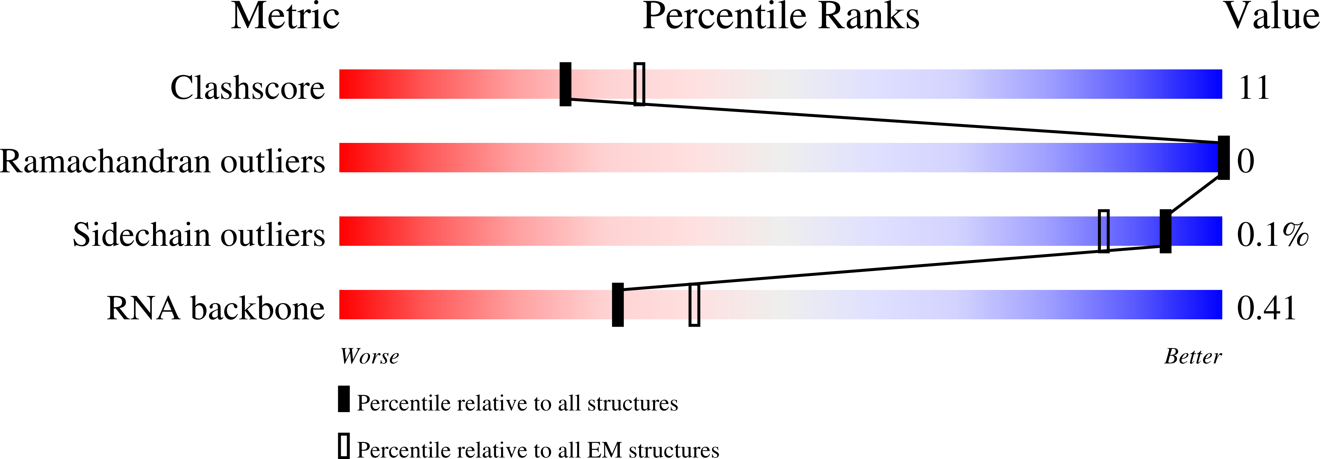
Method Details:
Experimental Method:
Resolution:
3.80 Å
Aggregation State:
PARTICLE
Reconstruction Method:
SINGLE PARTICLE