7LK4 image
Deposition Date 2021-02-01
Release Date 2022-02-09
Last Version Date 2024-11-06
Entry Detail
PDB ID:
7LK4
Keywords:
Title:
Crystal structure of BAK L100A in complex with activating antibody fragments
Biological Source:
Source Organism:
Homo sapiens (Taxon ID: 9606)
Rattus norvegicus (Taxon ID: 10116)
Host Organism:
Method Details:
Experimental Method:
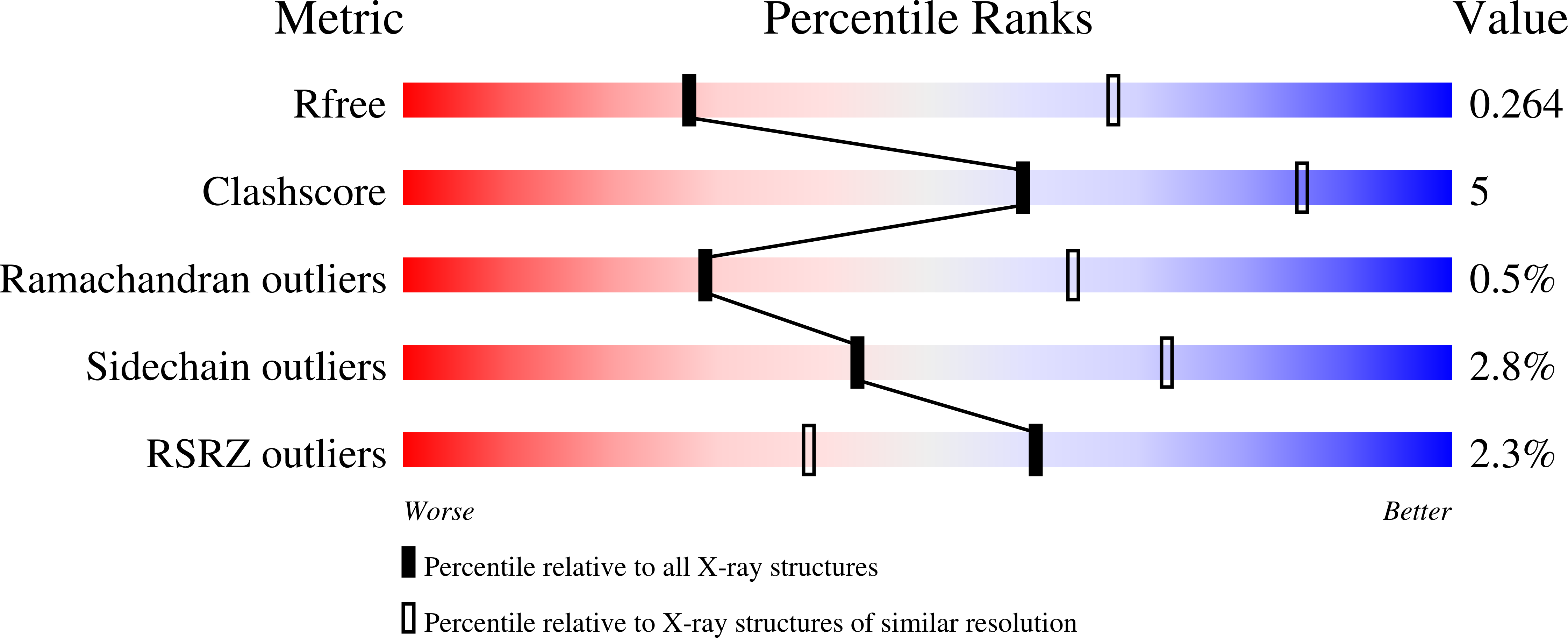
Resolution:
3.10 Å
R-Value Free:
0.26
R-Value Work:
0.21
R-Value Observed:
0.21
Space Group:
P 1 21 1
Macromolecular Entities
Polymer Type:polypeptide(L)
Molecule:7D10 antibody VL fragment
Chain IDs:E (auth: A), G (auth: C), I (auth: E), K (auth: G)
Chain Length:109
Number of Molecules:4
Biological Source:Rattus norvegicus
Polymer Type:polypeptide(L)
Molecule:7D10 antibody VH fragment
Chain IDs:F (auth: B), H (auth: D), J (auth: F), L (auth: H)
Chain Length:129
Number of Molecules:4
Biological Source:Rattus norvegicus
Polymer Type:polypeptide(L)
Molecule:Bcl-2 homologous antagonist/killer
Gene (Uniprot):BAK1
Mutations:L100A, C166S
Chain IDs:A (auth: P), B (auth: Q), C (auth: R), D (auth: S)
Chain Length:164
Number of Molecules:4
Biological Source:Homo sapiens
Ligand Molecules
Primary Citation
Structure of the BAK-activating antibody 7D10 bound to BAK reveals an unexpected role for the alpha 1-alpha 2 loop in BAK activation.
Cell Death Differ. 29 1757 1768 (2022)
PMID: 35279694 DOI: 10.1038/s41418-022-00961-w

Abstact

Pro-apoptotic BAK and BAX are activated by BH3-only proteins to permeabilise the outer mitochondrial membrane. The antibody 7D10 also activates BAK on mitochondria and its epitope has previously been mapped to BAK residues in the loop connecting helices α1 and α2 of BAK. A crystal structure of the complex between the Fv fragment of 7D10 and the BAK mutant L100A suggests a possible mechanism of activation involving the α1-α2 loop residue M60. M60 mutants of BAK have reduced stability and elevated sensitivity to activation by BID, illustrating that M60, through its contacts with residues in helices α1, α5 and α6, is a linchpin stabilising the inert, monomeric structure of BAK. Our data demonstrate that BAK's α1-α2 loop is not a passive covalent connector between secondary structure elements, but a direct restraint on BAK's activation.

Legend

Protein

Chemical

Disease

Primary Citation of related structures