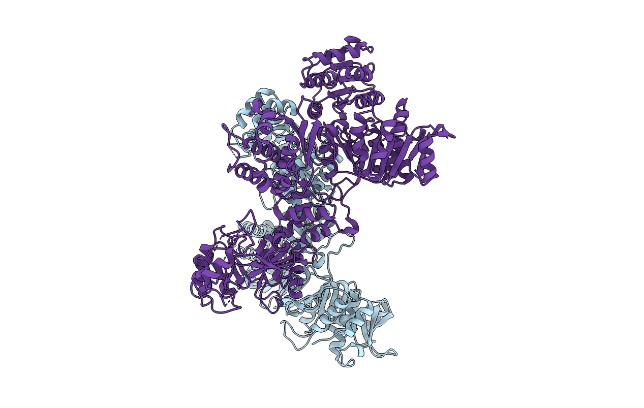
7LGN image
Entry Detail
PDB ID:
7LGN
Keywords:
Title:
Cyanophycin synthetase 1 from T. morbirosei
Biological Source:
Source Organism:
PDB Version:
Deposition Date:
2021-01-20
Release Date:
2021-08-18
Method Details:
Experimental Method:
Resolution:
3.10 Å
R-Value Free:
0.32
R-Value Work:
0.27
Space Group:
I 41
Macromolecular Entities
Polymer Type:polypeptide(L)
Description:Cyanophycin synthase
Chain IDs:A, B
Chain Length:909
Number of Molecules:2
Biological Source:Tatumella morbirosei
Ligand Molecules
Primary Citation
Structures and function of the amino acid polymerase cyanophycin synthetase.
Nat.Chem.Biol. 17 1101 1110 (2021)
PMID: 34385683 DOI: 10.1038/s41589-021-00854-y

Abstact

Cyanophycin is a natural biopolymer produced by a wide range of bacteria, consisting of a chain of poly-L-Asp residues with L-Arg residues attached to the β-carboxylate sidechains by isopeptide bonds. Cyanophycin is synthesized from ATP, aspartic acid and arginine by a homooligomeric enzyme called cyanophycin synthetase (CphA1). CphA1 has domains that are homologous to glutathione synthetases and muramyl ligases, but no other structural information has been available. Here, we present cryo-electron microscopy and X-ray crystallography structures of cyanophycin synthetases from three different bacteria, including cocomplex structures of CphA1 with ATP and cyanophycin polymer analogs at 2.6 Å resolution. These structures reveal two distinct tetrameric architectures, show the configuration of active sites and polymer-binding regions, indicate dynamic conformational changes and afford insight into catalytic mechanism. Accompanying biochemical interrogation of substrate binding sites, catalytic centers and oligomerization interfaces combine with the structures to provide a holistic understanding of cyanophycin biosynthesis.

Legend

Protein

Chemical

Disease

Primary Citation of related structures