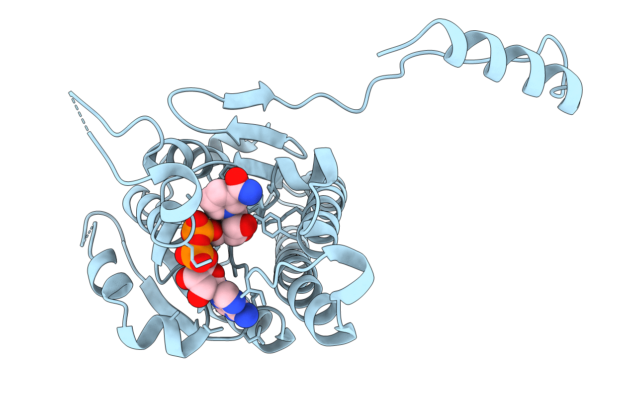
7LGB image
Entry Detail
PDB ID:
7LGB
Keywords:
Title:
ChsB1 in complex with NAD+
Biological Source:
PDB Version:
Deposition Date:
2021-01-19
Release Date:
2021-04-21
Method Details:
Experimental Method:
Resolution:
2.21 Å
R-Value Free:
0.20
R-Value Work:
0.16
R-Value Observed:
0.17
Space Group:
C 1 2 1
Macromolecular Entities
Polymer Type:polypeptide(L)
Description:3-ketoacyl-ACP reductase
Chain IDs:A
Chain Length:317
Number of Molecules:1
Biological Source:Mycobacterium tuberculosis
Ligand Molecules
Primary Citation
Enzymatic beta-Oxidation of the Cholesterol Side Chain in Mycobacterium tuberculosis Bifurcates Stereospecifically at Hydration of 3-Oxo-cholest-4,22-dien-24-oyl-CoA.
Acs Infect Dis. 7 1739 1751 (2021)
PMID: 33826843 DOI: 10.1021/acsinfecdis.1c00069

Abstact

The unique ability of Mycobacterium tuberculosis (Mtb) to utilize host lipids such as cholesterol for survival, persistence, and virulence has made the metabolic pathway of cholesterol an area of great interest for therapeutics development. Herein, we identify and characterize two genes from the Cho-region (genomic locus responsible for cholesterol catabolism) of the Mtb genome, chsH3 (Rv3538) and chsB1 (Rv3502c). Their protein products catalyze two sequential stereospecific hydration and dehydrogenation steps in the β-oxidation of the cholesterol side chain. ChsH3 favors the 22S hydration of 3-oxo-cholest-4,22-dien-24-oyl-CoA in contrast to the previously reported EchA19 (Rv3516), which catalyzes formation of the (22R)-hydroxy-3-oxo-cholest-4-en-24-oyl-CoA from the same enoyl-CoA substrate. ChsB1 is stereospecific and catalyzes dehydrogenation of the ChsH3 product but not the EchA19 product. The X-ray crystallographic structure of the ChsB1 apo-protein was determined at a resolution of 2.03 Å, and the holo-enzyme with bound NAD+ cofactor was determined at a resolution of 2.21 Å. The homodimeric structure is representative of a classical NAD+-utilizing short-chain type alcohol dehydrogenase/reductase, including a Rossmann-fold motif, but exhibits a unique substrate binding site architecture that is of greater length and width than its homologous counterparts, likely to accommodate the bulky steroid substrate. Intriguingly, Mtb utilizes hydratases from the MaoC-like family in sterol side-chain catabolism in contrast to fatty acid β-oxidation in other species that utilize the evolutionarily distinct crotonase family of hydratases.

Legend

Protein

Chemical

Disease

Primary Citation of related structures