Deposition Date
2021-01-12
Release Date
2021-09-01
Last Version Date
2024-10-30
Entry Detail
PDB ID:
7LD2
Keywords:
Title:
Zoogloea ramigera biosynthetic thiolase Q183Y mutant, RbCl soak
Biological Source:
Source Organism(s):
Zoogloea ramigera (Taxon ID: 350)
Expression System(s):
Method Details:
Experimental Method:
Resolution:
2.80 Å
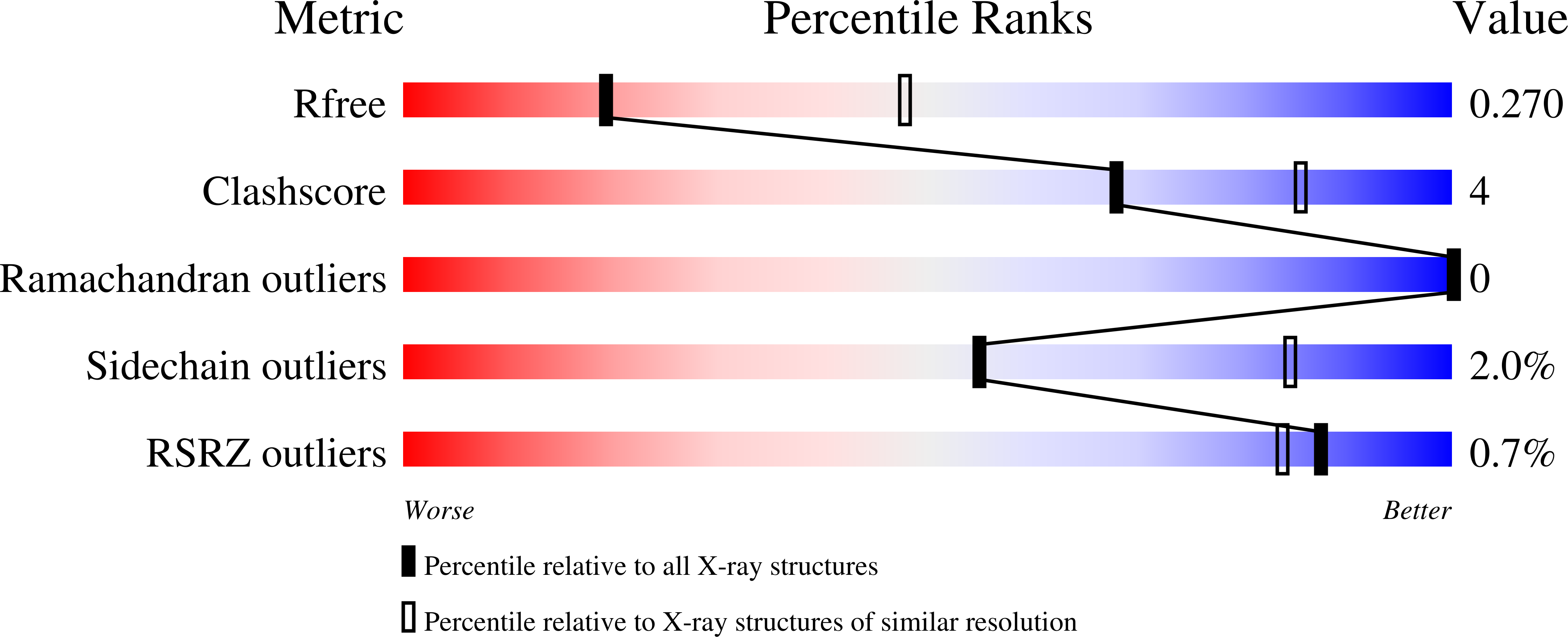
R-Value Free:
0.27
R-Value Work:
0.23
R-Value Observed:
0.23
Space Group:
P 1 21 1