Deposition Date
2021-01-11
Release Date
2021-09-01
Last Version Date
2024-11-06
Entry Detail
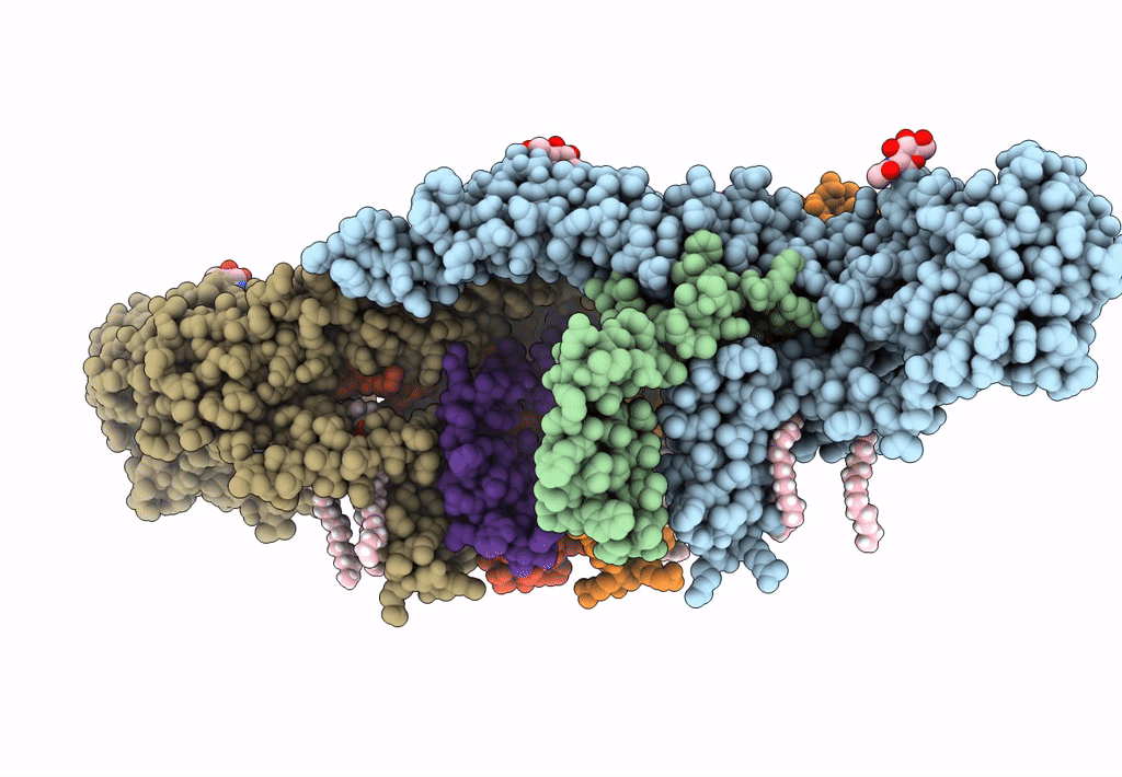
Biological Source:
Source Organism(s):
Usutu virus (Taxon ID: 64286)
Expression System(s):
Method Details:
Experimental Method:
Resolution:
2.35 Å
Aggregation State:
PARTICLE
Reconstruction Method:
SINGLE PARTICLE