Deposition Date
2020-12-24
Release Date
2021-12-22
Last Version Date
2024-11-06
Entry Detail
PDB ID:
7L6V
Keywords:
Title:
Crystal structure of BoNT/A-LC-JPU-A5-JPU-C1-JPU-H7-JPU-D12-ciA-F12
Biological Source:
Source Organism(s):
Clostridium botulinum (Taxon ID: 1491)
Vicugna pacos (Taxon ID: 30538)
Vicugna pacos (Taxon ID: 30538)
Expression System(s):
Method Details:
Experimental Method:
Resolution:
2.01 Å
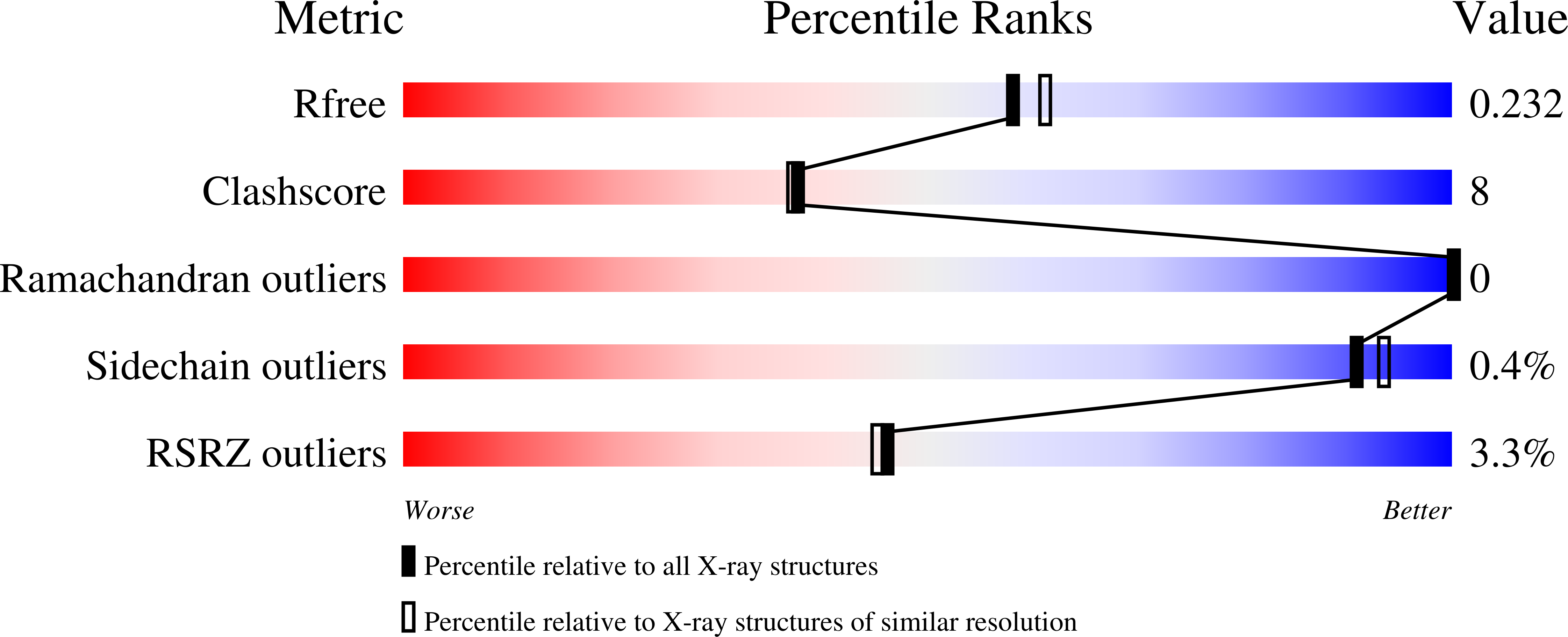
R-Value Free:
0.23
R-Value Work:
0.19
R-Value Observed:
0.19
Space Group:
I 2 2 2