7L0O image
Deposition Date 2020-12-11
Release Date 2021-09-08
Last Version Date 2024-10-09
Entry Detail
PDB ID:
7L0O
Keywords:
Title:
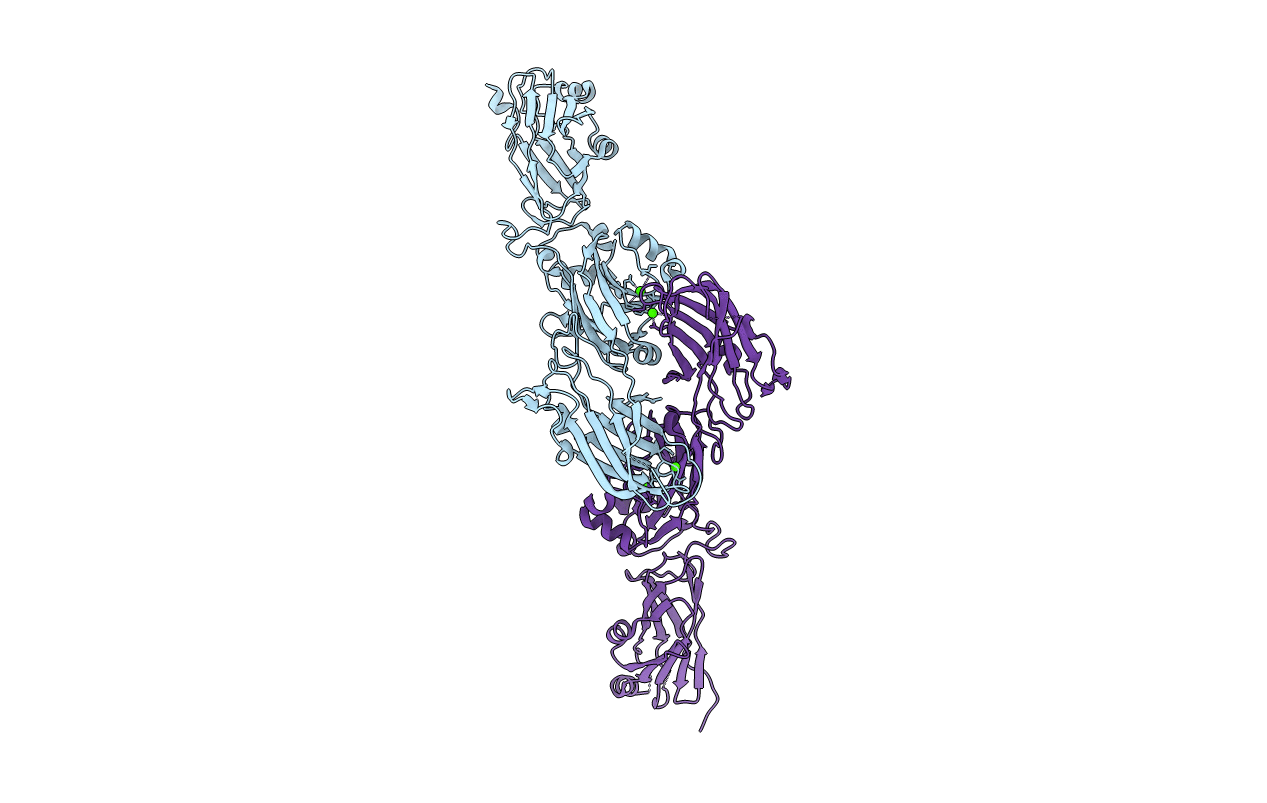
Streptococcus gordonii C123 Domain(s)-Structural and Functional Analysis
Biological Source:
Source Organism:
Method Details:
Experimental Method:
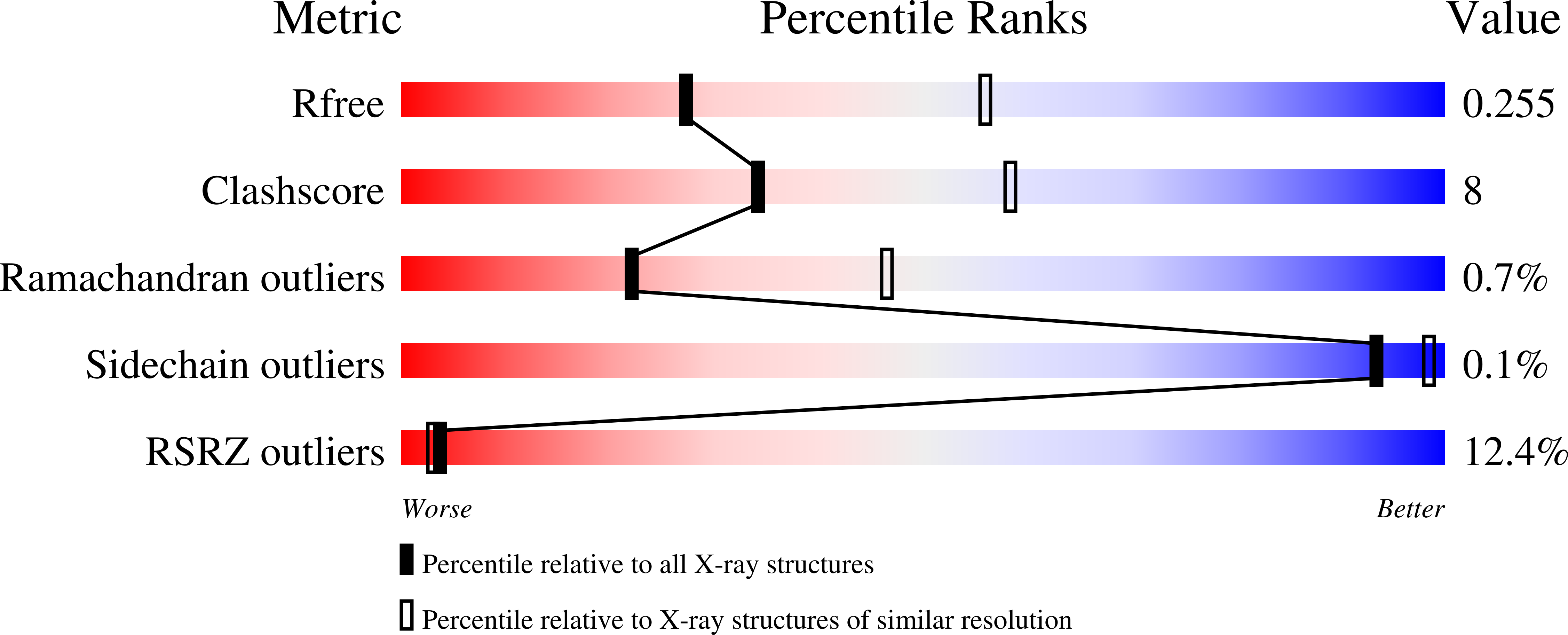
Resolution:
2.70 Å
R-Value Free:
0.25
R-Value Work:
0.21
R-Value Observed:
0.22
Space Group:
P 21 21 21
Macromolecular Entities
Structures with similar UniProt ID
Protein Blast
Polymer Type:polypeptide(L)
Molecule:Agglutinin receptor
Gene (Uniprot):sspB
Chain IDs:A, B
Chain Length:507
Number of Molecules:2
Biological Source:Streptococcus gordonii
Ligand Molecules
Primary Citation
Structural and functional analysis of the C-terminal region of Streptococcus gordonii SspB.
Acta Crystallogr D Struct Biol 77 1206 1215 (2021)
PMID: 34473090 DOI: 10.1107/S2059798321008135

Abstact

Streptococcus gordonii is a member of the viridans streptococci and is an early colonizer of the tooth surface. Adherence to the tooth surface is enabled by proteins present on the S. gordonii cell surface, among which SspB belongs to one of the most well studied cell-wall-anchored adhesin families: the antigen I/II (AgI/II) family. The C-terminal region of SspB consists of three tandemly connected individual domains that display the DEv-IgG fold. These C-terminal domains contain a conserved Ca2+-binding site and isopeptide bonds, and they adhere to glycoprotein 340 (Gp340; also known as salivary agglutinin, SAG). Here, the structural and functional characterization of the C123SspB domain at 2.7 Å resolution is reported. Although the individual C-terminal domains of Streptococcus mutans AgI/II and S. gordonii SspB show a high degree of both sequence and structural homology, superposition of these structures highlights substantial differences in their electrostatic surface plots, and this can be attributed to the relative orientation of the individual domains (C1, C2 and C3) with respect to each other and could reflect their specificity in binding to extracellular matrix molecules. Studies further confirmed that affinity for Gp340 or its scavenger receptor cysteine-rich (SRCR) domains requires two of the three domains of C123SspB, namely C12 or C23, which is different from AgI/II. Using protein-protein docking studies, models for this observed functional difference between C123SspB and C123AgI/II in their binding to SRCR1 are presented.

Legend

Protein

Chemical

Disease

Primary Citation of related structures
Feedback Form
Name
Email
Institute
Feedback