7KZ5 image
Deposition Date 2020-12-10
Release Date 2021-05-26
Last Version Date 2023-10-18
Entry Detail
PDB ID:
7KZ5
Title:
Crystal structure of KabA from Bacillus cereus UW85 in complex with the plp external aldimine adduct with kanosamine-6-phosphate
Biological Source:
Source Organism:
Bacillus cereus (Taxon ID: 1396)
Host Organism:
Method Details:
Experimental Method:
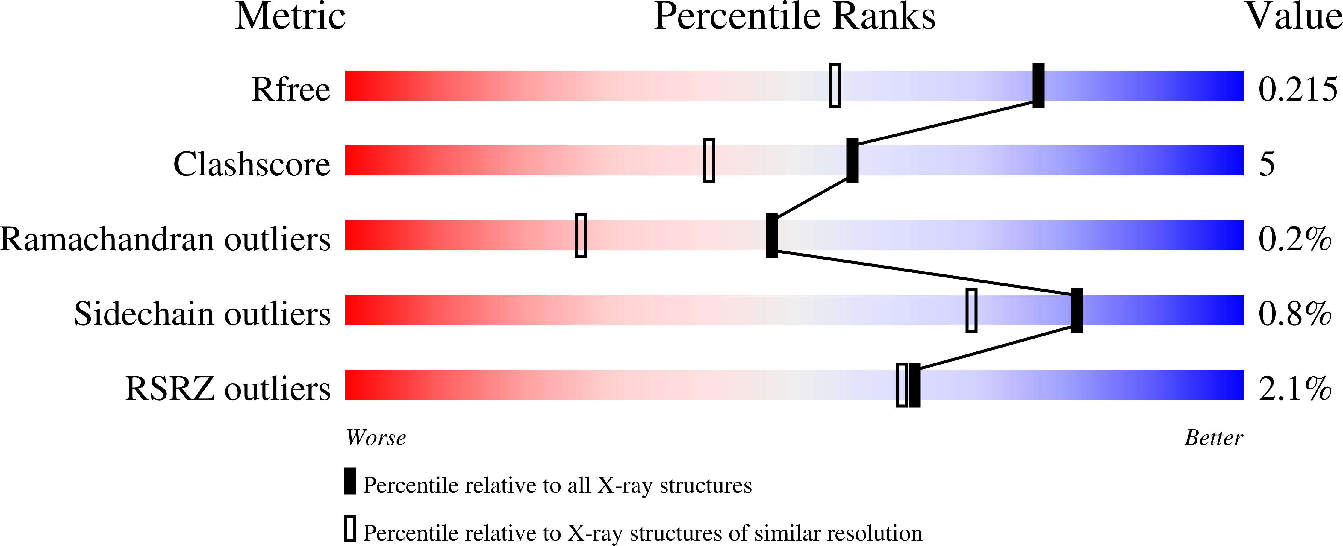
Resolution:
1.60 Å
R-Value Free:
0.21
R-Value Work:
0.17
R-Value Observed:
0.17
Space Group:
P 1
Macromolecular Entities
Structures with similar UniProt ID
Protein Blast
Polymer Type:polypeptide(L)
Molecule:Aminotransferase class I/II-fold pyridoxal phosphate-dependent enzyme
Gene (Uniprot):kabA
Chain IDs:A, B, C, D
Chain Length:445
Number of Molecules:4
Biological Source:Bacillus cereus
Primary Citation
Snapshots along the catalytic path of KabA, a PLP-dependent aminotransferase required for kanosamine biosynthesis in Bacillus cereus UW85.
J.Struct.Biol. 213 107744 107744 (2021)
PMID: 33984505 DOI: 10.1016/j.jsb.2021.107744

Abstact

Kanosamine is an antibiotic and antifungal monosaccharide. The kanosamine biosynthetic pathway from glucose 6-phosphate in Bacillus cereus UW85 was recently reported, and the functions of each of the three enzymes in the pathway, KabA, KabB and KabC, were demonstrated. KabA, a member of a subclass of the VIβ family of PLP-dependent aminotransferases, catalyzes the second step in the pathway, generating kanosamine 6-phosphate (K6P) using l-glutamate as the amino-donor. KabA catalysis was shown to be extremely efficient, with a second-order rate constant with respect to K6P transamination of over 107 M-1s-1. Here we report the high-resolution structure of KabA in both the PLP- and PMP-bound forms. In addition, co-crystallization with K6P allowed the structure of KabA in complex with the covalent PLP-K6P adduct to be solved. Co-crystallization or soaking with glutamate or 2-oxoglutarate did not result in crystals with either substrate/product. Reduction of the PLP-KabA complex with sodium cyanoborohydride gave an inactivated enzyme, and crystals of the reduced KabA were soaked with the l-glutamate analog glutarate to mimic the KabA-PLP-l-glutamate complex. Together these four structures give a complete picture of how the active site of KabA recognizes substrates for each half-reaction. The KabA structure is discussed in the context of homologous aminotransferases.

Legend

Protein

Chemical

Disease

Primary Citation of related structures