7KVT image
Entry Detail
PDB ID:
7KVT
Keywords:
Title:
Crystal structure of Squash RNA aptamer in complex with DFHBI-1T with iridium (III) ions
Biological Source:
Source Organism:
PDB Version:
Deposition Date:
2020-11-28
Release Date:
2022-01-19
Method Details:
Experimental Method:
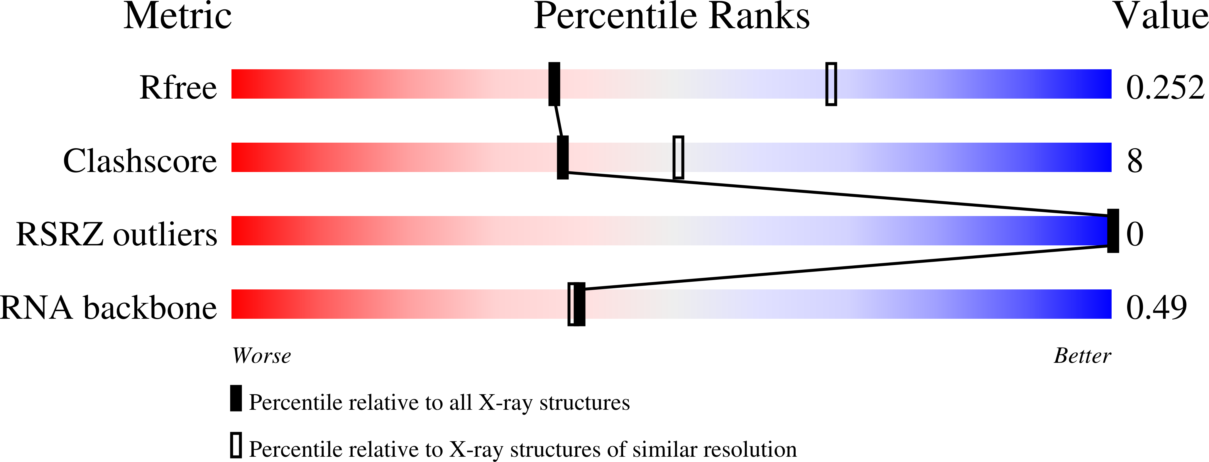
Resolution:
2.73 Å
R-Value Free:
0.25
R-Value Work:
0.20
R-Value Observed:
0.21
Space Group:
P 42 21 2
Macromolecular Entities
Polymer Type:polyribonucleotide
Description:Squash RNA aptamer bound to DFHBI-1T with iridium (III) ions
Chain IDs:A (auth: B)
Chain Length:83
Number of Molecules:1
Biological Source:synthetic construct
Primary Citation
The fluorescent aptamer Squash extensively repurposes the adenine riboswitch fold.
Nat.Chem.Biol. 18 191 198 (2022)
PMID: 34937911 DOI: 10.1038/s41589-021-00931-2

Abstact

Squash is an RNA aptamer that strongly activates the fluorescence of small-molecule analogs of the fluorophore of green fluorescent protein (GFP). Unlike other fluorogenic aptamers, isolated de novo from random-sequence RNA, Squash was evolved from the bacterial adenine riboswitch to leverage its optimized in vivo folding and stability. We now report the 2.7-Å resolution cocrystal structure of fluorophore-bound Squash, revealing that while the overall fold of the riboswitch is preserved, the architecture of the ligand-binding core is dramatically transformed. Unlike previously characterized aptamers that activate GFP-derived fluorophores, Squash does not harbor a G-quadruplex, sandwiching its fluorophore between a base triple and a noncanonical base quadruple in a largely apolar pocket. The expanded structural core of Squash allows it to recognize unnatural fluorophores that are larger than the simple purine ligand of the parental adenine riboswitch, and suggests that stable RNA scaffolds can tolerate larger variation than has hitherto been appreciated.

Legend

Protein

Chemical

Disease

Primary Citation of related structures