7KV1 image
Deposition Date 2020-11-26
Release Date 2021-02-17
Last Version Date 2024-03-06
Entry Detail
PDB ID:
7KV1
Title:
Surface glycan-binding protein A from Bacteroides uniformis
Biological Source:
Method Details:
Experimental Method:
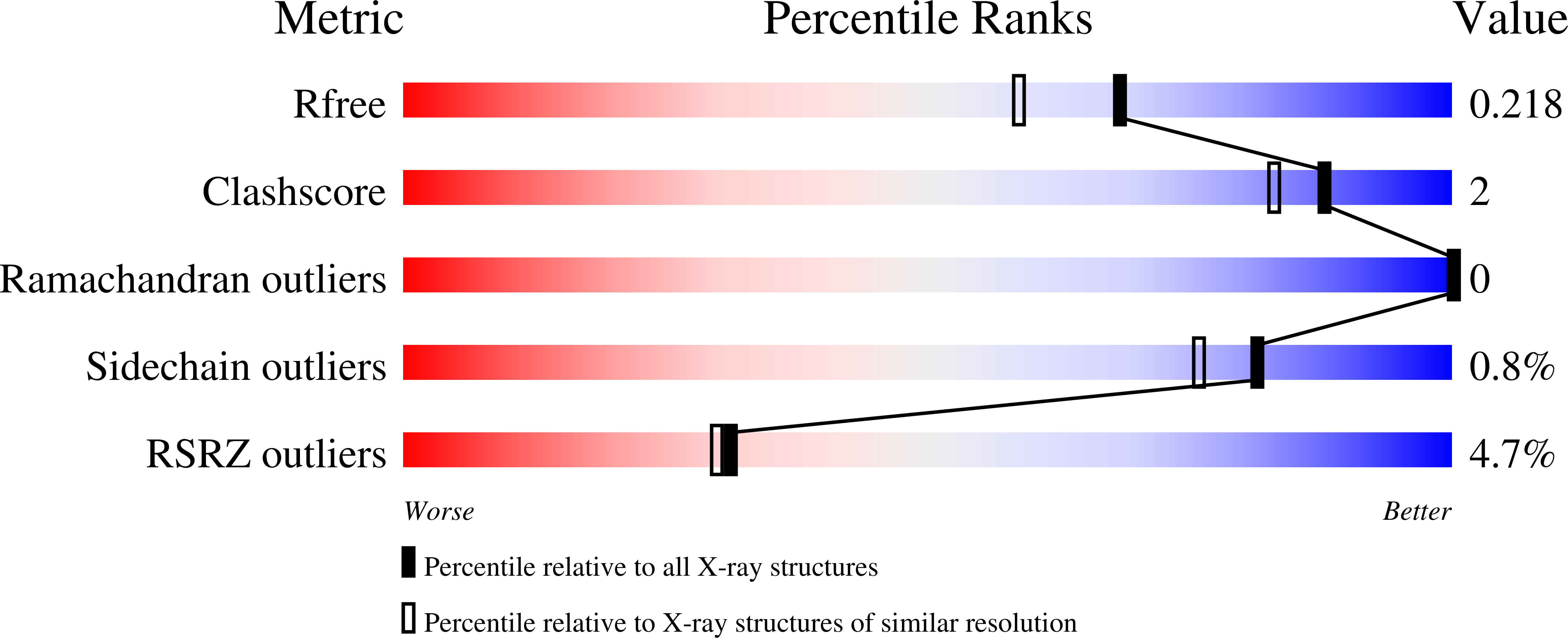
Resolution:
1.86 Å
R-Value Free:
0.20
R-Value Work:
0.16
R-Value Observed:
0.16
Space Group:
P 1 21 1
Macromolecular Entities
Polymer Type:polypeptide(L)
Molecule:SusD family protein
Chain IDs:A, B
Chain Length:529
Number of Molecules:2
Biological Source:Bacteroides uniformis (strain ATCC 8492 / DSM 6597 / CIP 103695 / JCM 5828 / NCTC 13054 / VPI 0061)
Primary Citation
Distinct protein architectures mediate species-specific beta-glucan binding and metabolism in the human gut microbiota.
J.Biol.Chem. 296 100415 100415 (2021)
PMID: 33587952 DOI: 10.1016/j.jbc.2021.100415

Abstact

Complex glycans that evade our digestive system are major nutrients that feed the human gut microbiota (HGM). The prevalence of Bacteroidetes in the HGM of populations worldwide is engendered by the evolution of polysaccharide utilization loci (PULs), which encode concerted protein systems to utilize the myriad complex glycans in our diets. Despite their crucial roles in glycan recognition and transport, cell-surface glycan-binding proteins (SGBPs) remained understudied cogs in the PUL machinery. Here, we report the structural and biochemical characterization of a suite of SGBP-A and SGBP-B structures from three syntenic β(1,3)-glucan utilization loci (1,3GULs) from Bacteroides thetaiotaomicron (Bt), Bacteroides uniformis (Bu), and B. fluxus (Bf), which have varying specificities for distinct β-glucans. Ligand complexes provide definitive insight into β(1,3)-glucan selectivity in the HGM, including structural features enabling dual β(1,3)-glucan/mixed-linkage β(1,3)/β(1,4)-glucan-binding capability in some orthologs. The tertiary structural conservation of SusD-like SGBPs-A is juxtaposed with the diverse architectures and binding modes of the SGBPs-B. Specifically, the structures of the trimodular BtSGBP-B and BuSGBP-B revealed a tandem repeat of carbohydrate-binding module-like domains connected by long linkers. In contrast, BfSGBP-B comprises a bimodular architecture with a distinct β-barrel domain at the C terminus that bears a shallow binding canyon. The molecular insights obtained here contribute to our fundamental understanding of HGM function, which in turn may inform tailored microbial intervention therapies.

Legend

Protein

Chemical

Disease

Primary Citation of related structures