Deposition Date
2020-11-24
Release Date
2021-06-30
Last Version Date
2023-10-18
Entry Detail
PDB ID:
7KUF
Keywords:
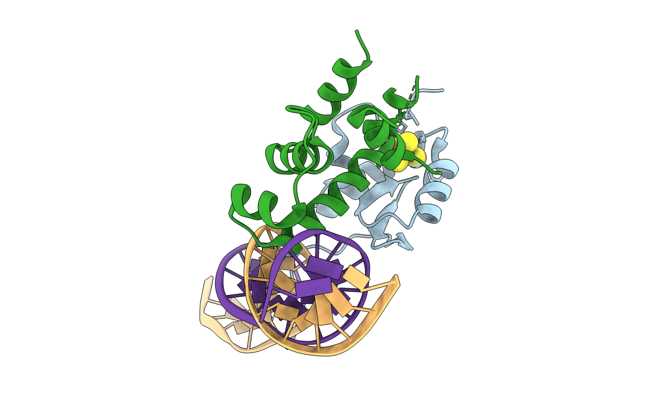
Title:
Transcription activation subcomplex with WhiB7 bound to SigmaAr4-RNAP Beta flap tip chimera and DNA
Biological Source:
Source Organism(s):
Mycobacterium tuberculosis (Taxon ID: 1773)
Expression System(s):
Method Details:
Experimental Method:
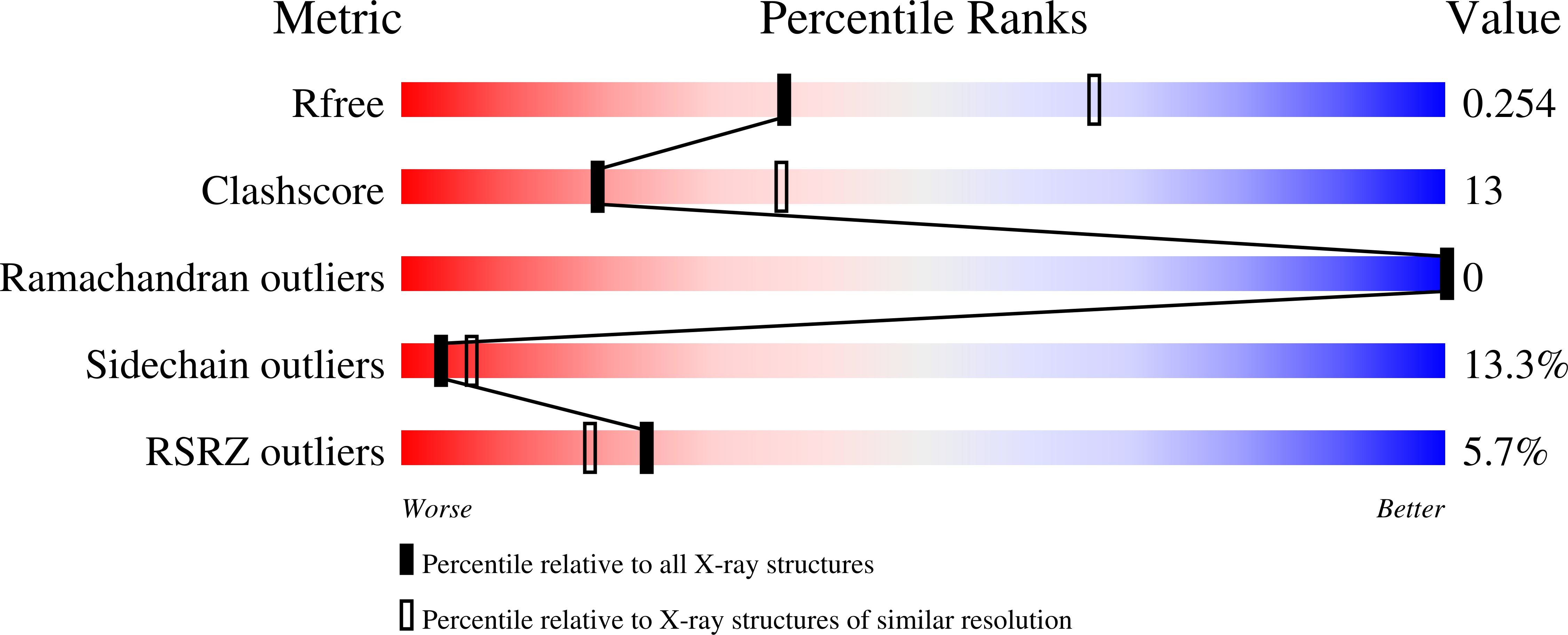
Resolution:
2.60 Å
R-Value Free:
0.25
R-Value Work:
0.22
R-Value Observed:
0.22
Space Group:
P 32 2 1