7KQT image
Entry Detail
PDB ID:
7KQT
Keywords:
Title:
A 1.84-A resolution crystal structure of heme-dependent L-tyrosine hydroxylase in complex with 3-fluoro-L-tyrosine and cyanide
Biological Source:
Source Organism:
Host Organism:
PDB Version:
Deposition Date:
2020-11-17
Release Date:
2021-03-31
Method Details:
Experimental Method:
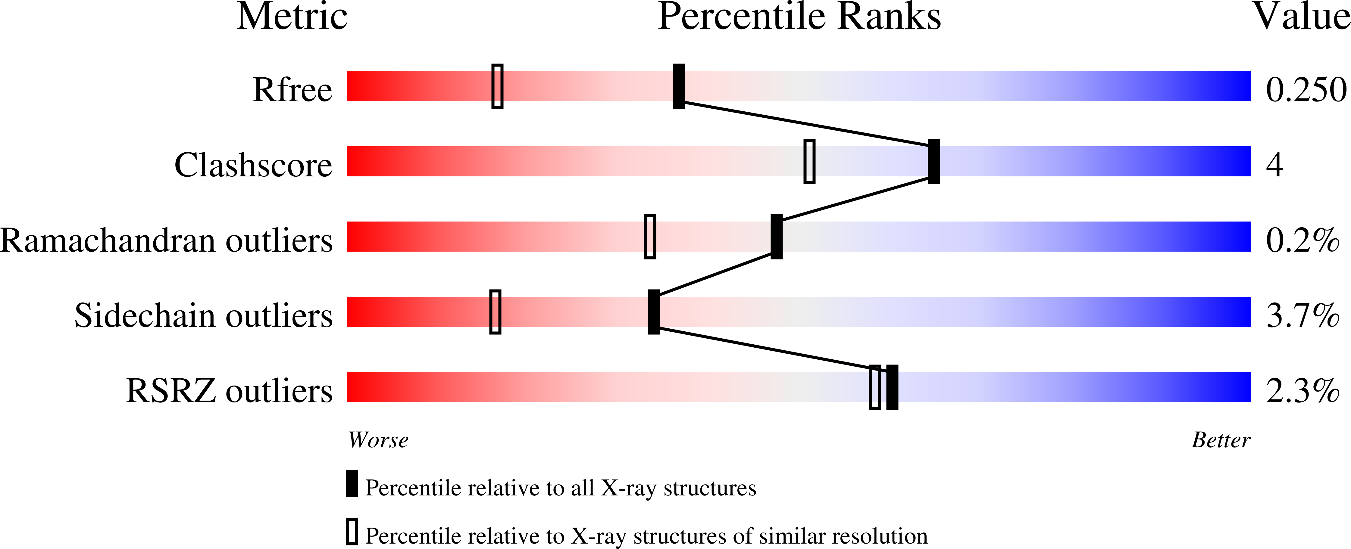
Resolution:
1.84 Å
R-Value Free:
0.25
R-Value Work:
0.20
R-Value Observed:
0.20
Space Group:
P 1 21 1
Macromolecular Entities
Polymer Type:polypeptide(L)
Description:Heme-dependent L-tyrosine hydroxylase
Chain IDs:A, B
Chain Length:316
Number of Molecules:2
Biological Source:Streptomyces sclerotialus
Primary Citation
Molecular Rationale for Partitioning between C-H and C-F Bond Activation in Heme-Dependent Tyrosine Hydroxylase.
J.Am.Chem.Soc. 143 4680 4693 (2021)
PMID: 33734681 DOI: 10.1021/jacs.1c00175

Abstact

The heme-dependent l-tyrosine hydroxylases (TyrHs) in natural product biosynthesis constitute a new enzyme family in contrast to the nonheme iron enzymes for DOPA production. A representative TyrH exhibits dual reactivity of C-H and C-F bond cleavage when challenged with 3-fluoro-l-tyrosine (3-F-Tyr) as a substrate. However, little is known about how the enzyme mediates two distinct reactions. Herein, a new TyrH from the thermophilic bacterium Streptomyces sclerotialus (SsTyrH) was functionally and structurally characterized. A de novo crystal structure of the enzyme-substrate complex at 1.89-Å resolution provides the first comprehensive structural study of this hydroxylase. The binding conformation of l-tyrosine indicates that C-H bond hydroxylation is initiated by electron transfer. Mutagenesis studies confirmed that an active site histidine, His88, participates in catalysis. We also obtained a 1.68-Å resolution crystal structure in complex with the monofluorinated substrate, 3-F-Tyr, which shows one binding conformation but two orientations of the fluorine atom with a ratio of 7:3, revealing that the primary factor of product distribution is the substrate orientation. During in crystallo reaction, a ferric-hydroperoxo intermediate (compound 0, Fe3+-OOH) was observed with 3-F-Tyr as a substrate based on characteristic spectroscopic features. We determined the crystal structure of this compound 0-type intermediate and refined it to 1.58-Å resolution. Collectively, this study provided the first molecular details of the heme-dependent TyrH and determined the primary factor that dictates the partitioning between the dual reactivities of C-H and C-F bond activation.

Legend

Protein

Chemical

Disease

Primary Citation of related structures