Deposition Date
2020-10-26
Release Date
2021-04-07
Last Version Date
2023-10-18
Entry Detail
PDB ID:
7KJZ
Keywords:
Title:
crystal structure of PLEKHA7 PH domain biding inositol-tetraphosphate
Biological Source:
Source Organism(s):
Homo sapiens (Taxon ID: 9606)
Expression System(s):
Method Details:
Experimental Method:
Resolution:
2.43 Å
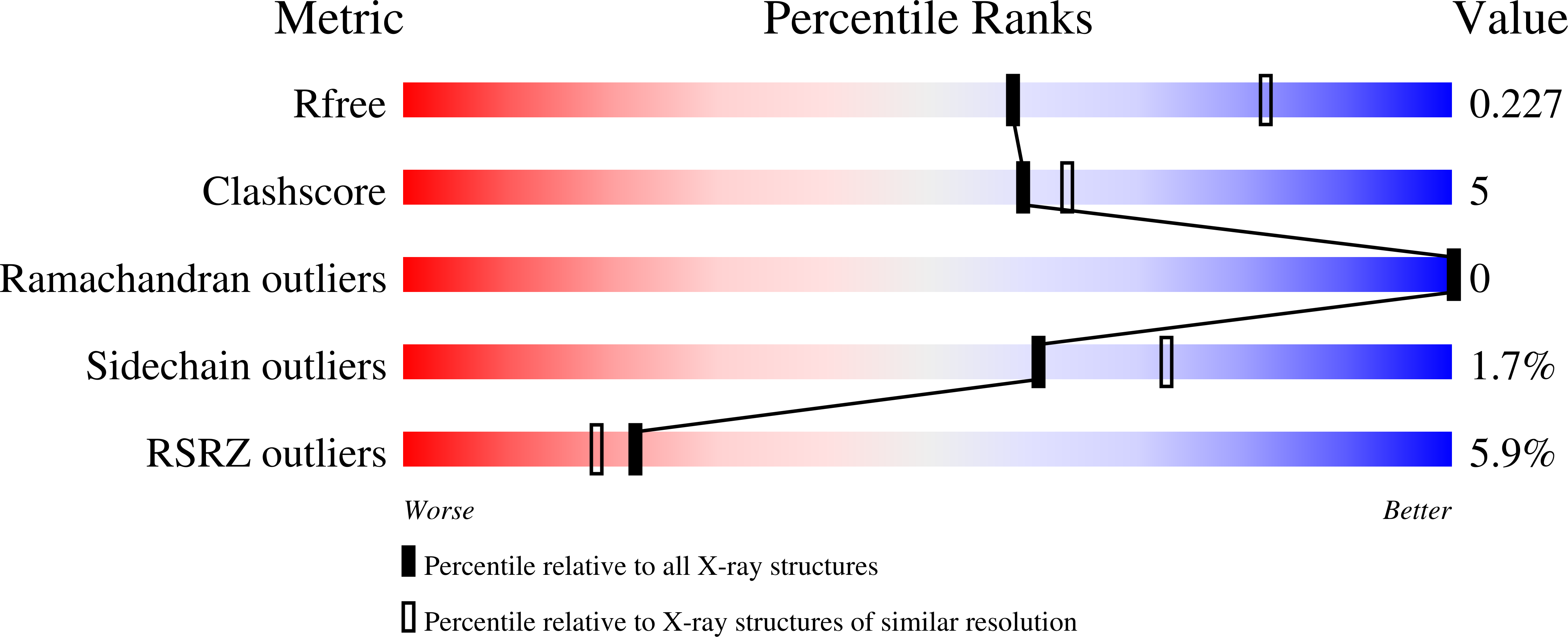
R-Value Free:
0.22
R-Value Work:
0.18
R-Value Observed:
0.18
Space Group:
P 32 2 1