Deposition Date
2020-10-26
Release Date
2021-05-19
Last Version Date
2023-11-15
Entry Detail
PDB ID:
7KJJ
Keywords:
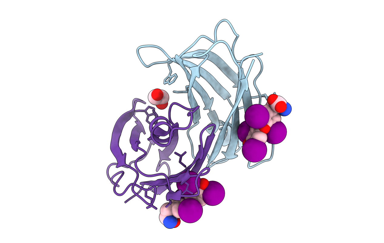
Title:
Reconstructed ancestor of HIUases and Transthyretins
Biological Source:
Source Organism(s):
unidentified (Taxon ID: 32644)
Expression System(s):
Method Details:
Experimental Method:
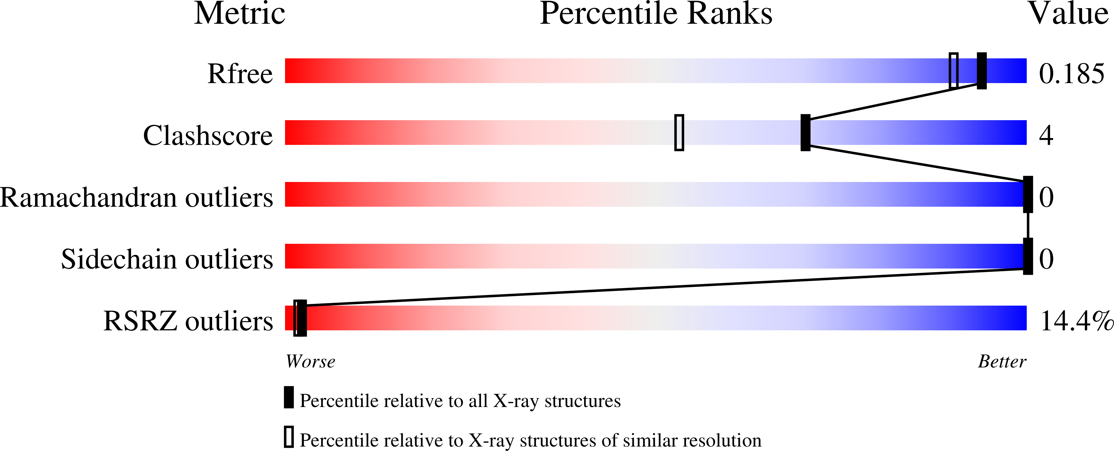
Resolution:
1.55 Å
R-Value Free:
0.18
R-Value Work:
0.17
R-Value Observed:
0.17
Space Group:
P 43 21 2