Deposition Date
2020-10-23
Release Date
2021-07-07
Last Version Date
2023-10-18
Entry Detail
PDB ID:
7KIL
Keywords:
Title:
Crystal structure of the mouse lipin-1 M-Lip domain with zinc
Biological Source:
Source Organism(s):
Mus musculus (Taxon ID: 10090)
Expression System(s):
Method Details:
Experimental Method:
Resolution:
1.90 Å
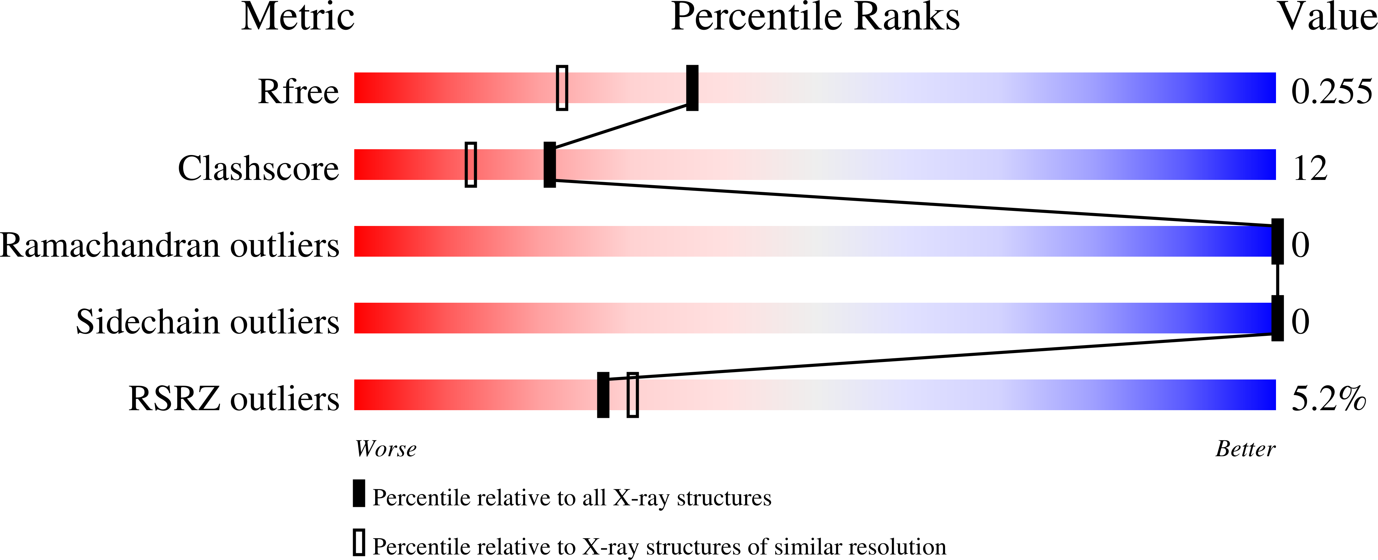
R-Value Free:
0.25
R-Value Work:
0.22
R-Value Observed:
0.22
Space Group:
P 1 21 1