7KHT image
Deposition Date 2020-10-22
Release Date 2021-05-19
Last Version Date 2023-10-18
Entry Detail
PDB ID:
7KHT
Title:
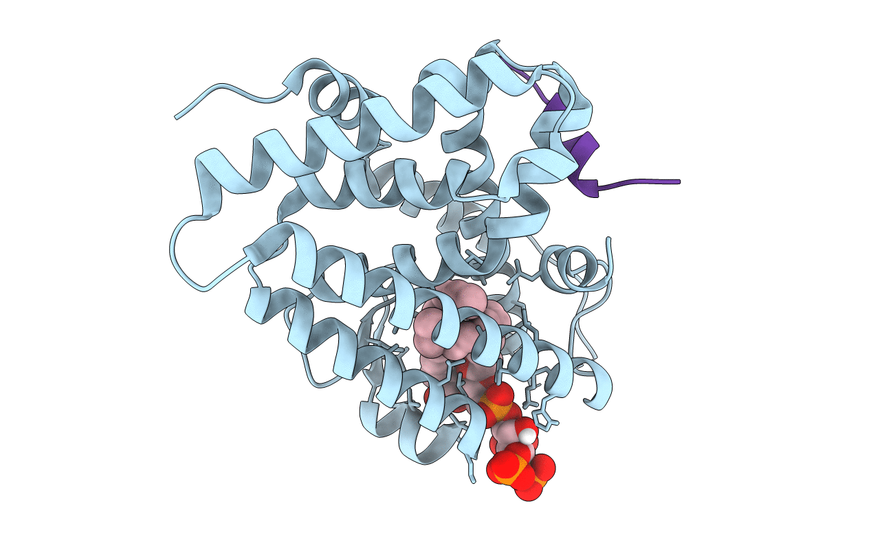
The acyl chains of phosphoinositide PIP3 alter the structure and function of nuclear receptor Steroidogenic Factor-1 (SF-1)
Biological Source:
Source Organism:
Homo sapiens (Taxon ID: 9606)
Host Organism:
Method Details:
Experimental Method:
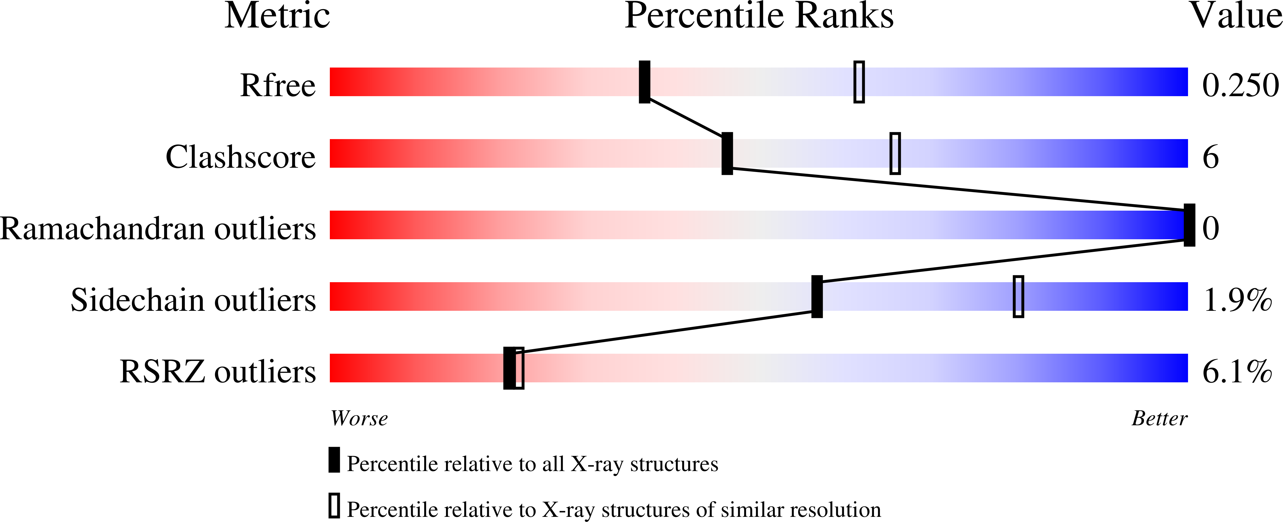
Resolution:
2.50 Å
R-Value Free:
0.24
R-Value Work:
0.21
R-Value Observed:
0.22
Space Group:
P 41 21 2
Macromolecular Entities
Polymer Type:polypeptide(L)
Molecule:Steroidogenic factor 1
Gene (Uniprot):NR5A1
Mutations:C247S, C412S
Chain IDs:A
Chain Length:245
Number of Molecules:1
Biological Source:Homo sapiens
Polymer Type:polypeptide(L)
Molecule:Peroxisome proliferator-activated receptor gamma coactivator 1-alpha peptide
Gene (Uniprot):PPARGC1A
Chain IDs:B
Chain Length:14
Number of Molecules:1
Biological Source:Homo sapiens
Ligand Molecules
Primary Citation
The acyl chains of phosphoinositide PIP3 alter the structure and function of nuclear receptor steroidogenic factor-1.
J.Lipid Res. 62 100081 100081 (2021)
PMID: 33933440 DOI: 10.1016/j.jlr.2021.100081

Abstact

Nuclear receptors are transcription factors that bind lipids, an event that induces a structural conformation of the receptor that favors interaction with transcriptional coactivators. The nuclear receptor steroidogenic factor-1 (SF-1, NR5A1) binds the signaling phosphoinositides PI(4,5)P2 (PIP2) and PI(3,4,5)P3 (PIP3), and our previous crystal structures showed how the phosphoinositide headgroups regulate SF-1 function. However, what role the acyl chains play in regulating SF-1 structure remains unaddressed. Here, we used X-ray crystallography with in vitro binding and functional assays to examine how the acyl chains of PIP3 regulate human SF-1 ligand-binding domain structure and function. Altering acyl chain length and unsaturation regulates apparent binding of all tested phosphoinositides to SF-1. Mass spectrometry-based lipidomics data suggest C16 and C18 phospholipids preferentially associate with SF-1 expressed ectopically in bacteria. We then solved the 2.5 Å crystal structure of SF-1 bound to dioleoyl PIP3(18:1/18:1) to compare it with a matched structure of SF-1 bound to dipalmitoyl PIP3(16:0/16:0). The dioleoyl-bound structure was severely disordered in a specific SF-1 region associated with pathogenic human polymorphisms and within the coactivator-binding region critical for SF-1 function while inducing increased sensitivity to protease digestion in solution. Validating these structural observations, in vitro functional studies showed dioleoyl PIP3 induced 6-fold poorer affinity of a peroxisome proliferator-activated receptor gamma coactivator 1-alpha coactivator peptide for SF-1 compared with dipalmitoyl PIP3. Together, these data suggest the chemical nature of the phosphoinositide acyl chains controls the ordered state of specific, clinically important structural regions in SF-1, regulating SF-1 function in vitro.

Legend

Protein

Chemical

Disease

Primary Citation of related structures