7KD5 image
Entry Detail
PDB ID:
7KD5
Keywords:
Title:
Structure of the C-terminal domain of the Menangle virus phosphoprotein (residues 329 -388), fused to MBP. Space group P212121
Biological Source:
PDB Version:
Deposition Date:
2020-10-08
Release Date:
2021-10-06
Method Details:
Experimental Method:
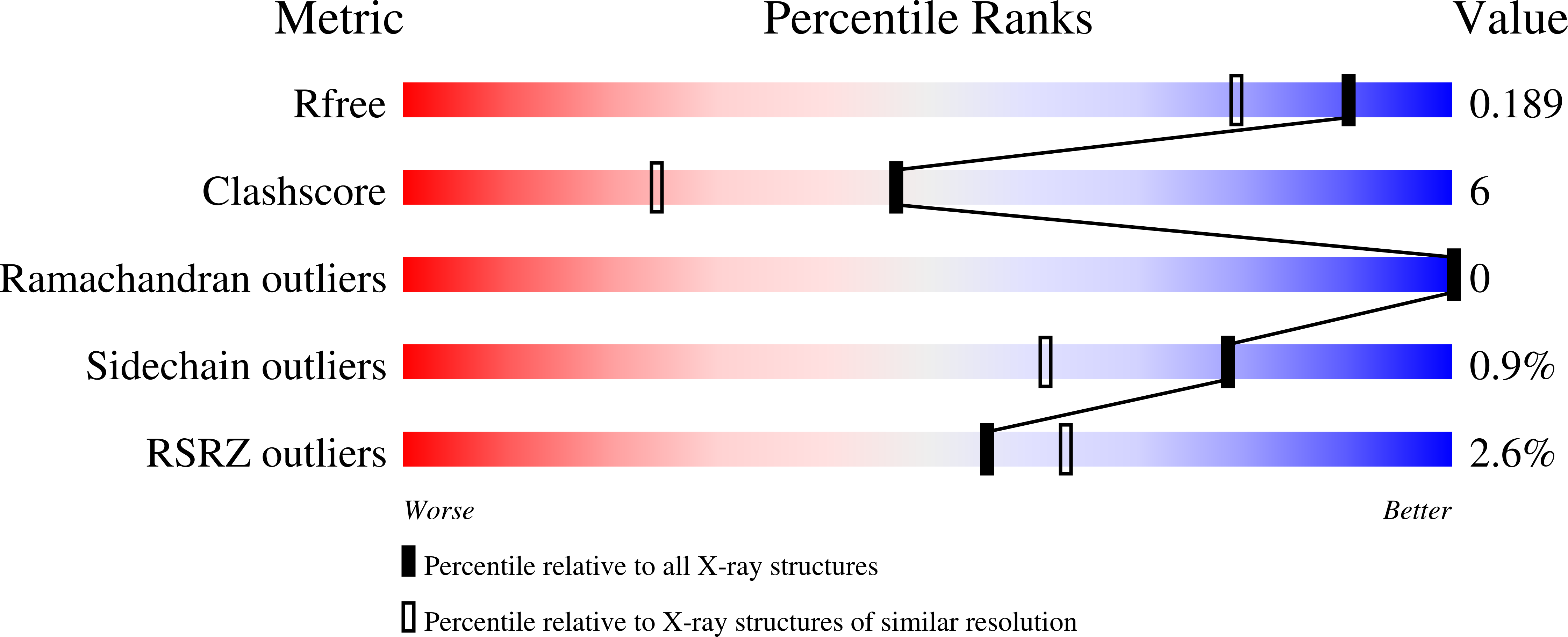
Resolution:
1.55 Å
R-Value Free:
0.18
R-Value Work:
0.16
Space Group:
P 21 21 21
Macromolecular Entities
Polymer Type:polypeptide(L)
Description:Maltodextrin-binding protein and Phosphoprotein fusion protein
Mutations:C352S
Chain IDs:A
Chain Length:431
Number of Molecules:1
Biological Source:Serratia sp. (strain FS14), Menangle virus
Peptide-like Molecules
PRD_900001
Primary Citation
Structural Analysis of the Menangle Virus P Protein Reveals a Soft Boundary between Ordered and Disordered Regions.
Viruses 13 ? ? (2021)
PMID: 34578318 DOI: 10.3390/v13091737

Abstact

The paramyxoviral phosphoprotein (P protein) is the non-catalytic subunit of the viral RNA polymerase, and coordinates many of the molecular interactions required for RNA synthesis. All paramyxoviral P proteins oligomerize via a centrally located coiled-coil that is connected to a downstream binding domain by a dynamic linker. The C-terminal region of the P protein coordinates interactions between the catalytic subunit of the polymerase, and the viral nucleocapsid housing the genomic RNA. The inherent flexibility of the linker is believed to facilitate polymerase translocation. Here we report biophysical and structural characterization of the C-terminal region of the P protein from Menangle virus (MenV), a bat-borne paramyxovirus with zoonotic potential. The MenV P protein is tetrameric but can dissociate into dimers at sub-micromolar protein concentrations. The linker is globally disordered and can be modeled effectively as a worm-like chain. However, NMR analysis suggests very weak local preferences for alpha-helical and extended beta conformation exist within the linker. At the interface between the disordered linker and the structured C-terminal binding domain, a gradual disorder-to-order transition occurs, with X-ray crystallographic analysis revealing a dynamic interfacial structure that wraps the surface of the binding domain.

Legend

Protein

Chemical

Disease

Primary Citation of related structures