7K7Y image
Entry Detail
PDB ID:
7K7Y
Keywords:
Title:
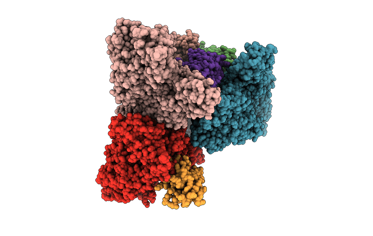
Crystal structure of BoNT/E LC-HN domain in complex with VHH JLE-E9
Biological Source:
Host Organism:
PDB Version:
Deposition Date:
2020-09-24
Release Date:
2020-10-14
Method Details:
Experimental Method:
Resolution:
3.60 Å
R-Value Free:
0.28
R-Value Work:
0.26
R-Value Observed:
0.26
Space Group:
P 21 21 21
Macromolecular Entities
Polymer Type:polypeptide(L)
Description:Botulinum neurotoxin type E
Chain IDs:A, C (auth: E), D (auth: G), E (auth: B)
Chain Length:850
Number of Molecules:4
Biological Source:Clostridium botulinum
Polymer Type:polypeptide(L)
Description:JLE-E9
Chain IDs:B (auth: D), F (auth: C), G (auth: F), H
Chain Length:129
Number of Molecules:4
Biological Source:Vicugna pacos
Ligand Molecules
Primary Citation
Two VHH Antibodies Neutralize Botulinum Neurotoxin E1 by Blocking Its Membrane Translocation in Host Cells.
Toxins 12 ? ? (2020)
PMID: 32992561 DOI: 10.3390/toxins12100616

Abstact

Botulinum neurotoxin serotype E (BoNT/E) is one of the major causes of human botulism, which is a life-threatening disease caused by flaccid paralysis of muscles. After receptor-mediated toxin internalization into motor neurons, the translocation domain (HN) of BoNT/E transforms into a protein channel upon vesicle acidification in endosomes and delivers its protease domain (LC) across membrane to enter the neuronal cytosol. It is believed that the rapid onset of BoNT/E intoxication compared to other BoNT serotypes is related to its swift internalization and translocation. We recently identified two neutralizing single-domain camelid antibodies (VHHs) against BoNT/E1 termed JLE-E5 and JLE-E9. Here, we report the crystal structures of these two VHHs bound to the LCHN domain of BoNT/E1. The structures reveal that these VHHs recognize two distinct epitopes that are partially overlapping with the putative transmembrane regions on HN, and therefore could physically block membrane association of BoNT/E1. This is confirmed by our in vitro studies, which show that these VHHs inhibit the structural change of BoNT/E1 at acidic pH and interfere with BoNT/E1 association with lipid vesicles. Therefore, these two VHHs neutralize BoNT/E1 by preventing the transmembrane delivery of LC. Furthermore, structure-based sequence analyses show that the 3-dimensional epitopes of these two VHHs are largely conserved across many BoNT/E subtypes, suggesting a broad-spectrum protection against the BoNT/E family. In summary, this work improves our understanding of the membrane translocation mechanism of BoNT/E and paves the way for developing VHHs as diagnostics or therapeutics for the treatment of BoNT/E intoxication.

Legend

Protein

Chemical

Disease

Primary Citation of related structures