Deposition Date
2020-09-10
Release Date
2021-03-17
Last Version Date
2023-10-18
Entry Detail
PDB ID:
7K32
Keywords:
Title:
Crystal structure of Endonuclease Q complex with 27-mer duplex substrate with an abasic lesion at the active site
Biological Source:
Source Organism(s):
Pyrococcus furiosus (Taxon ID: 2261)
synthetic construct (Taxon ID: 32630)
synthetic construct (Taxon ID: 32630)
Expression System(s):
Method Details:
Experimental Method:
Resolution:
3.11 Å
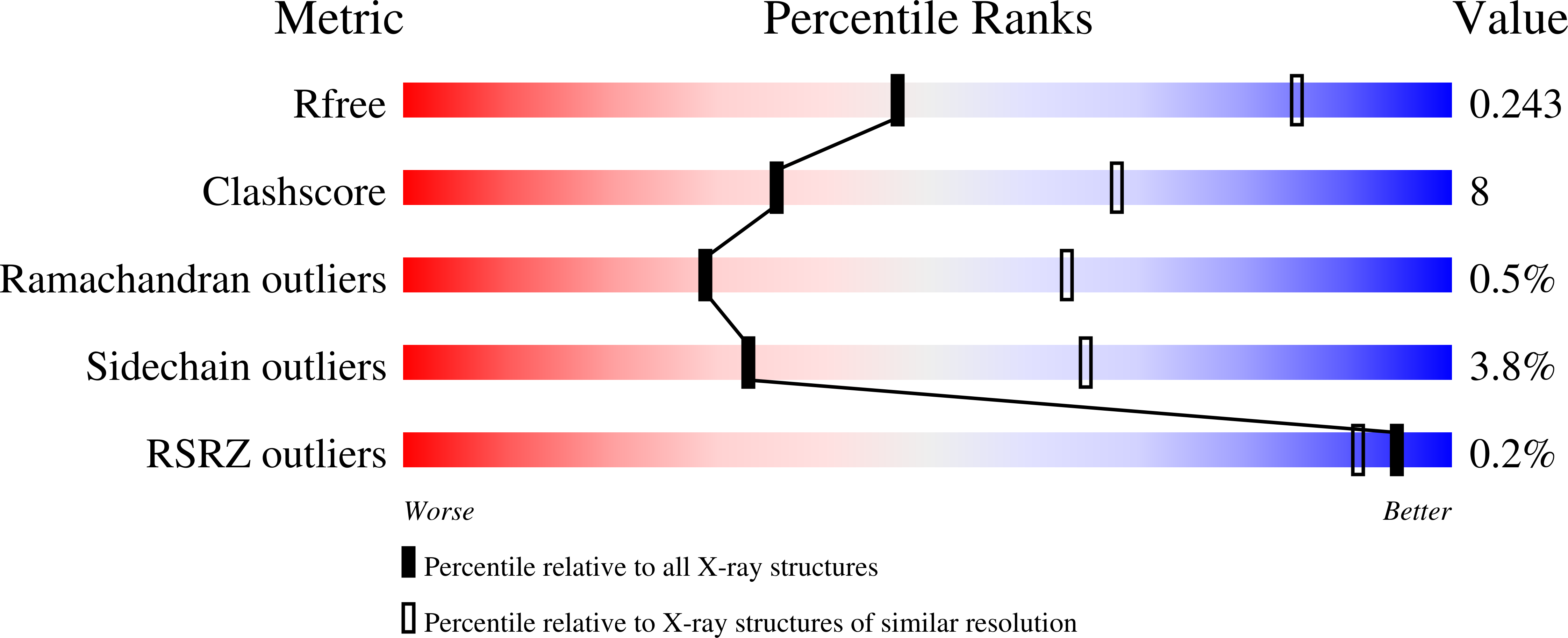
R-Value Free:
0.24
R-Value Work:
0.19
R-Value Observed:
0.20
Space Group:
H 3