Deposition Date
2020-08-17
Release Date
2021-09-01
Last Version Date
2024-10-30
Entry Detail
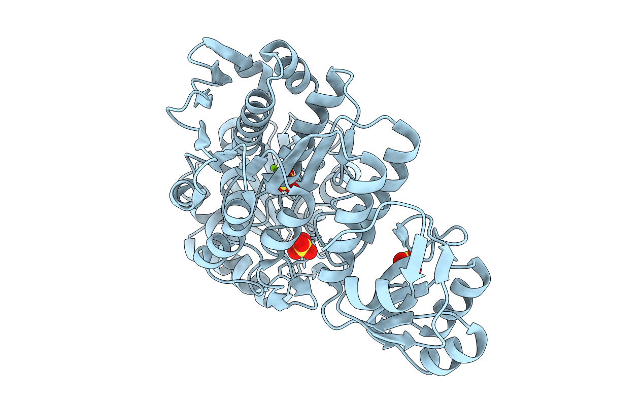
PDB ID:
7JT8
Keywords:
Title:
Apo structure of a pseudomurein peptide ligase type E from Methanothermus fervidus
Biological Source:
Source Organism(s):
Expression System(s):
Method Details:
Experimental Method:
Resolution:
1.84 Å
R-Value Free:
0.21
R-Value Work:
0.17
R-Value Observed:
0.17
Space Group:
P 61