Deposition Date
2020-08-15
Release Date
2020-10-21
Last Version Date
2025-05-14
Entry Detail
PDB ID:
7JSN
Keywords:
Title:
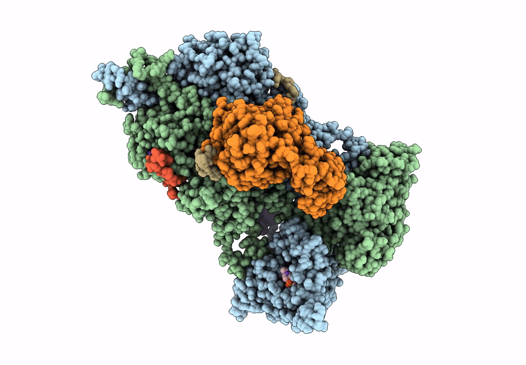
Structure of the Visual Signaling Complex between Transducin and Phosphodiesterase 6
Biological Source:
Source Organism(s):
Bos taurus (Taxon ID: 9913)
Expression System(s):
Method Details:
Experimental Method:
Resolution:
3.20 Å
Aggregation State:
PARTICLE
Reconstruction Method:
SINGLE PARTICLE