7JRQ image
Deposition Date 2020-08-12
Release Date 2021-01-13
Last Version Date 2023-10-18
Entry Detail
PDB ID:
7JRQ
Keywords:
Title:
Crystallographically Characterized De Novo Designed Mn-Diphenylporphyrin Binding Protein
Biological Source:
Source Organism:
Escherichia coli (Taxon ID: 562)
Host Organism:
Method Details:
Experimental Method:
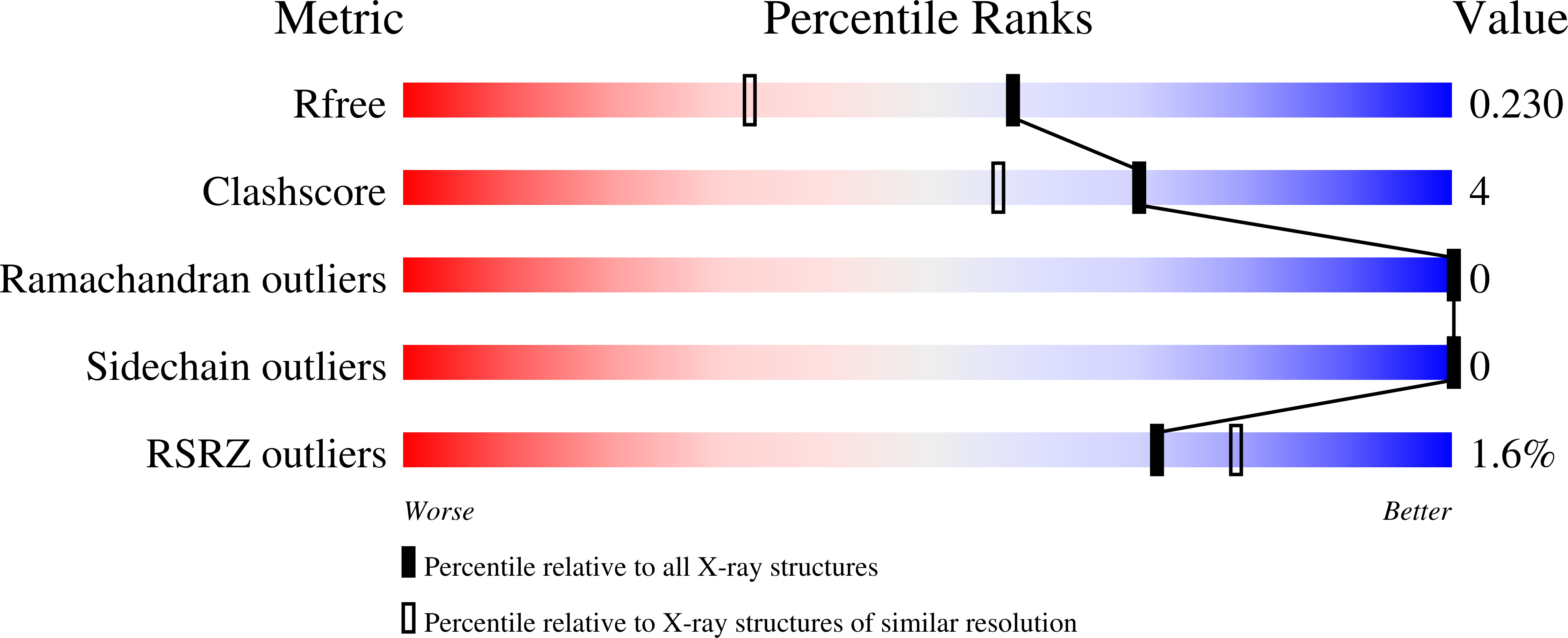
Resolution:
1.75 Å
R-Value Free:
0.22
R-Value Work:
0.19
R-Value Observed:
0.19
Space Group:
P 41 21 2
Macromolecular Entities
Polymer Type:polypeptide(L)
Molecule:MPP1
Chain IDs:A
Chain Length:131
Number of Molecules:1
Biological Source:Escherichia coli
Primary Citation
De Novo Design, Solution Characterization, and Crystallographic Structure of an Abiological Mn-Porphyrin-Binding Protein Capable of Stabilizing a Mn(V) Species.
J.Am.Chem.Soc. 143 252 259 (2021)
PMID: 33373215 DOI: 10.1021/jacs.0c10136

Abstact

De novo protein design offers the opportunity to test our understanding of how metalloproteins perform difficult transformations. Attaining high-resolution structural information is critical to understanding how such designs function. There have been many successes in the design of porphyrin-binding proteins; however, crystallographic characterization has been elusive, limiting what can be learned from such studies as well as the extension to new functions. Moreover, formation of highly oxidizing high-valent intermediates poses design challenges that have not been previously implemented: (1) purposeful design of substrate/oxidant access to the binding site and (2) limiting deleterious oxidation of the protein scaffold. Here we report the first crystallographically characterized porphyrin-binding protein that was programmed to not only bind a synthetic Mn-porphyrin but also maintain binding site access to form high-valent oxidation states. We explicitly designed a binding site with accessibility to dioxygen units in the open coordination site of the Mn center. In solution, the protein is capable of accessing a high-valent Mn(V)-oxo species which can transfer an O atom to a thioether substrate. The crystallographic structure is within 0.6 Å of the design and indeed contained an aquo ligand with a second water molecule stabilized by hydrogen bonding to a Gln side chain in the active site, offering a structural explanation for the observed reactivity.

Legend

Protein

Chemical

Disease

Primary Citation of related structures