7JID image
Deposition Date 2020-07-23
Release Date 2020-08-26
Last Version Date 2023-10-18
Entry Detail
PDB ID:
7JID
Keywords:
Title:
Crystal structure of the L780 UDP-rhamnose synthase from Acanthamoeba polyphaga mimivirus
Biological Source:
Source Organism:
Host Organism:
Method Details:
Experimental Method:
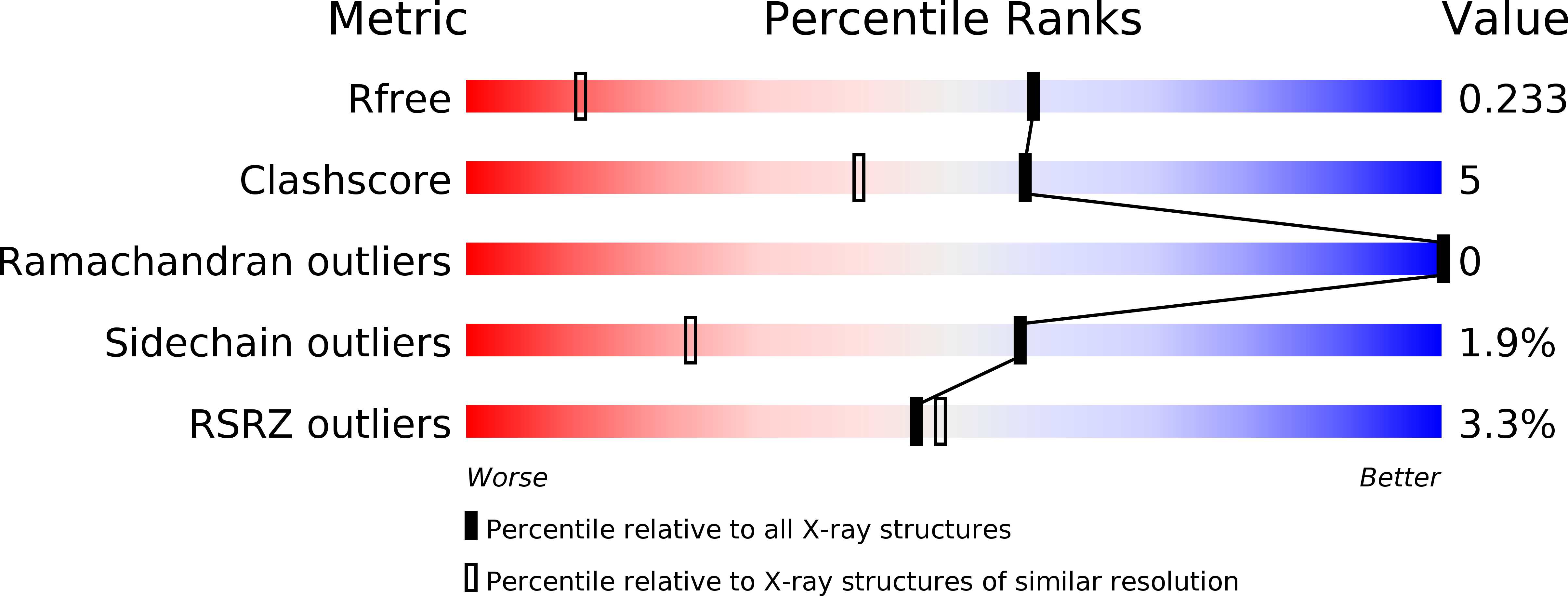
Resolution:
1.45 Å
R-Value Free:
0.22
R-Value Work:
0.19
R-Value Observed:
0.19
Space Group:
P 1
Macromolecular Entities
Polymer Type:polypeptide(L)
Molecule:UDP-L-Rhamnose Synthase
Gene (Uniprot):MIMI_L780
Chain IDs:A, B
Chain Length:292
Number of Molecules:2
Biological Source:Acanthamoeba polyphaga mimivirus
Primary Citation
The high-resolution structure of a UDP-L-rhamnose synthase from Acanthamoeba polyphaga Mimivirus.
Protein Sci. 29 2164 2174 (2020)
PMID: 32797646 DOI: 10.1002/pro.3928

Abstact

For the field of virology, perhaps one of the most paradigm-shifting events so far in the 21st century was the identification of the giant double-stranded DNA virus that infects amoebae. Remarkably, this virus, known as Mimivirus, has a genome that encodes for nearly 1,000 proteins, some of which are involved in the biosynthesis of unusual sugars. Indeed, the virus is coated by a layer of glycosylated fibers that contain d-glucose, N-acetyl-d-glucosamine, l-rhamnose, and 4-amino-4,6-dideoxy-d-glucose. Here we describe a combined structural and enzymological investigation of the protein encoded by the open-reading frame L780, which corresponds to an l-rhamnose synthase. The structure of the L780/NADP+ /UDP-l-rhamnose ternary complex was determined to 1.45 Å resolution and refined to an overall R-factor of 19.9%. Each subunit of the dimeric protein adopts a bilobal-shaped appearance with the N-terminal domain harboring the dinucleotide-binding site and the C-terminal domain positioning the UDP-sugar into the active site. The overall molecular architecture of L780 places it into the short-chain dehydrogenase/reductase superfamily. Kinetic analyses indicate that the enzyme can function on either UDP- and dTDP-sugars but displays a higher catalytic efficiency with the UDP-linked substrate. Site-directed mutagenesis experiments suggest that both Cys 108 and Lys 175 play key roles in catalysis. This structure represents the first model of a viral UDP-l-rhamnose synthase and provides new details into these fascinating enzymes.

Legend

Protein

Chemical

Disease

Primary Citation of related structures