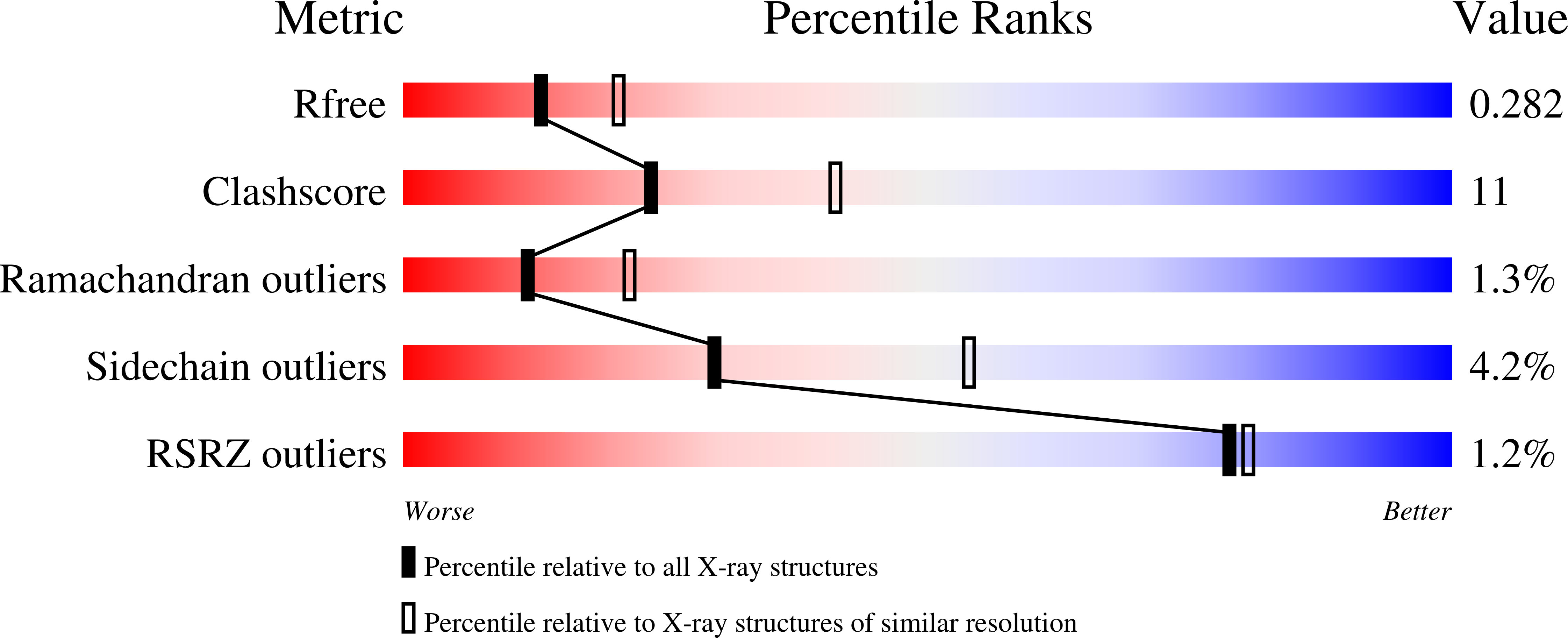
7JH0 image
Deposition Date 2020-07-20
Release Date 2021-02-17
Last Version Date 2024-11-20
Entry Detail
PDB ID:
7JH0
Keywords:
Title:
Crystallographic structure of glyceraldehyde-3-phosphate dehydrogenase from Schistosoma mansoni
Biological Source:
Source Organism:
Host Organism:
Method Details:
Experimental Method:
Resolution:
2.51 Å
R-Value Free:
0.27
R-Value Work:
0.21
R-Value Observed:
0.21
Space Group:
P 61
Macromolecular Entities
Polymer Type:polypeptide(L)
Molecule:Glyceraldehyde-3-phosphate dehydrogenase
Chain IDs:A, B, C
Chain Length:338
Number of Molecules:3
Biological Source:Schistosoma mansoni
Polymer Type:polypeptide(L)
Molecule:Glyceraldehyde-3-phosphate dehydrogenase
Chain IDs:D
Chain Length:338
Number of Molecules:1
Biological Source:Schistosoma mansoni
Modified Residue
Compound ID Chain ID Parent Comp ID Details 2D Image
CSD D CYS modified residue
CSP A CYS modified residue
Primary Citation
Structure determination and analyses of the GAPDH from the parasite Schistosoma mansoni, the first one from a platyhelminth.
Biochimie 184 18 25 (2021)
PMID: 33524435 DOI: 10.1016/j.biochi.2021.01.014

Abstact

The enzyme Glyceraldehyde-3-Phosphate Dehydrogenase from Schistosoma mansoni (SmGAPDH) is characterized as a therapeutical target for schistosomiasis. In this context, we report here the experimental structure, structural analyses and comparisons of SmGAPDH, the first one from a Platyhelminth. The enzyme was expressed, purified and assayed for crystallization, what allowed the obtainment of crystals of sufficient quality to collect X-ray diffraction data up to 2.51 Å resolution. SmGAPDH is the only GAPDH to present the sequence NNR (its residues 114-116) which leads to (especially R116) a hydrogen bond network that possibly reflects on the flexibility of residues to interact with the adenine part of NAD+, speculated to be important for differential drug design.

Legend

Protein

Chemical

Disease

Primary Citation of related structures