Deposition Date
2024-09-16
Release Date
2025-08-20
Last Version Date
2025-08-20
Entry Detail
PDB ID:
7HI2
Keywords:
Title:
PanDDA analysis group deposition -- Crystal structure of SARS-CoV-2 NSP3 macrodomain in complex with AVI-0000682
Biological Source:
Source Organism(s):
Expression System(s):
Method Details:
Experimental Method:
Resolution:
1.03 Å
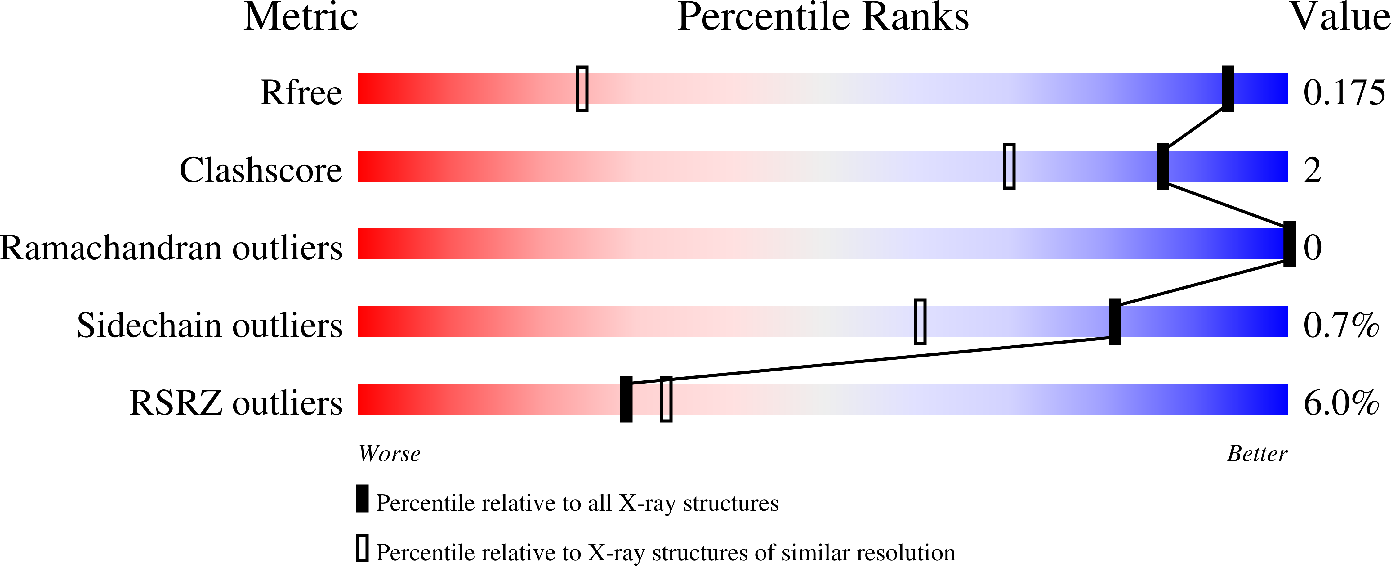
R-Value Free:
0.17
R-Value Work:
0.15
R-Value Observed:
0.15
Space Group:
P 43