
Deposition Date
2024-04-04
Release Date
2024-04-24
Last Version Date
2024-10-16
Entry Detail
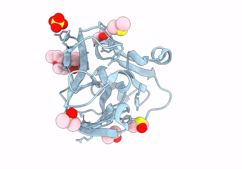
PDB ID:
7H2U
Keywords:
Title:
Group deposition for crystallographic fragment screening of Coxsackievirus A16 (G-10) 2A protease -- Crystal structure of Coxsackievirus A16 (G-10) 2A protease in complex with Z355202286 (A71EV2A-x0188)
Biological Source:
Source Organism(s):
Coxsackievirus A16 (Taxon ID: 31704)
Expression System(s):
Method Details:
Experimental Method:
Resolution:
1.11 Å
R-Value Free:
0.19
R-Value Work:
0.18
R-Value Observed:
0.18
Space Group:
C 1 2 1
