Deposition Date
2021-07-26
Release Date
2021-09-08
Last Version Date
2023-11-29
Entry Detail
PDB ID:
7FG6
Keywords:
Title:
Crystal structure of the Tyrosyl-tRNA synthetase (TyrRS) in Nanoarchaeum equitans
Biological Source:
Source Organism(s):
Nanoarchaeum equitans Kin4-M (Taxon ID: 228908)
Expression System(s):
Method Details:
Experimental Method:
Resolution:
2.80 Å
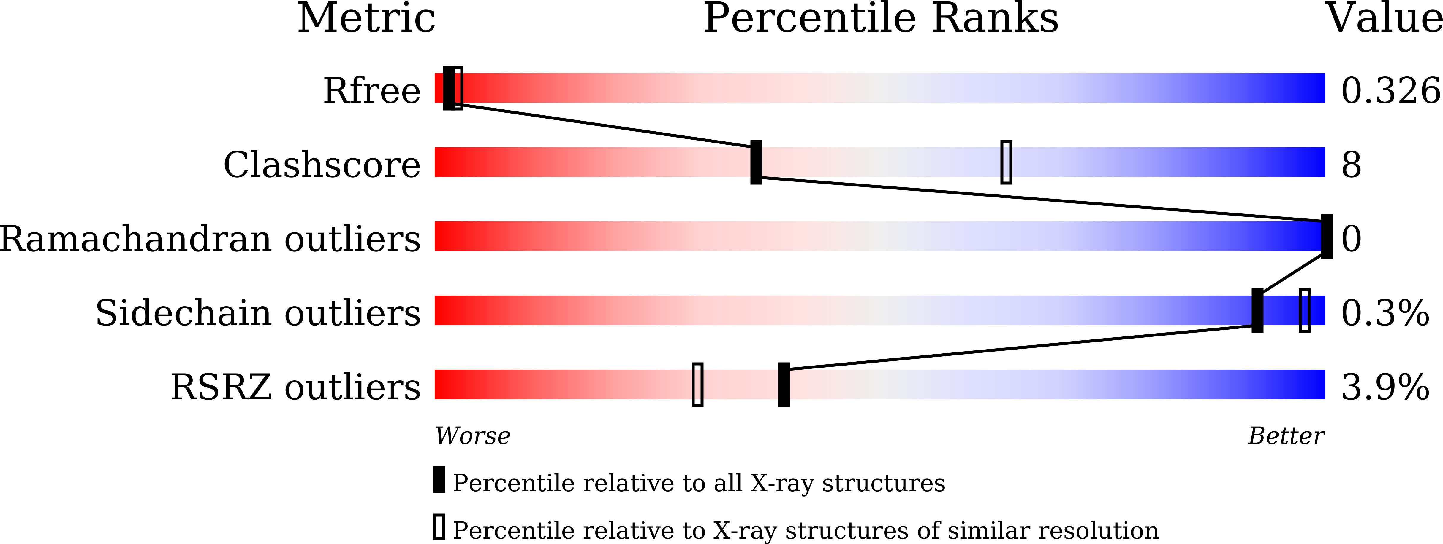
R-Value Free:
0.32
R-Value Work:
0.28
R-Value Observed:
0.28
Space Group:
P 32 2 1