7FDW image
Entry Detail
PDB ID:
7FDW
Keywords:
Title:
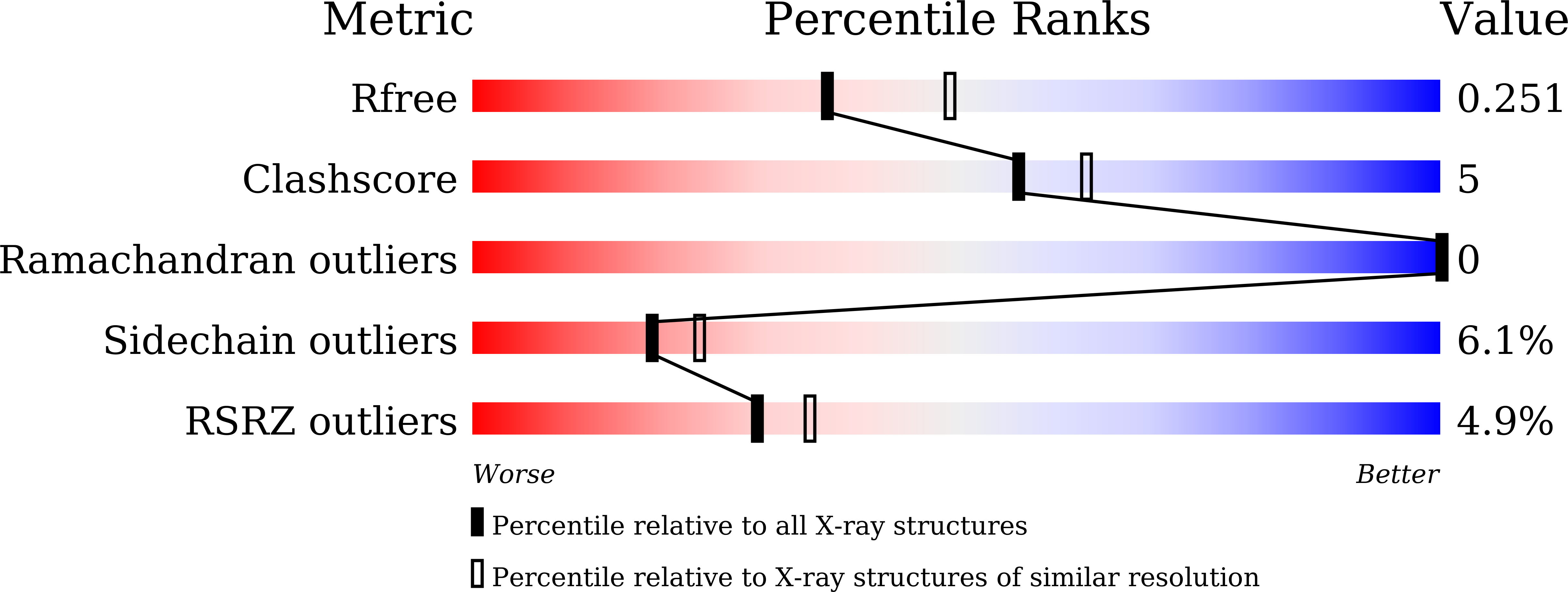
Crystal structure of pepsin cleaved lactoferrin C-lobe at 2.28 A resolution
Biological Source:
Source Organism:
PDB Version:
Deposition Date:
2021-07-18
Release Date:
2021-08-04
Method Details:
Experimental Method:
Resolution:
2.28 Å
R-Value Free:
0.24
R-Value Work:
0.21
Space Group:
C 1 2 1
Macromolecular Entities
Polymer Type:polypeptide(L)
Description:Lactotransferrin
Chain IDs:A, B
Chain Length:348
Number of Molecules:2
Biological Source:Bos taurus
Primary Citation
A Peptide Bond from the Inter-lobe Segment in the Bilobal Lactoferrin Acts as a Preferred Site for Cleavage for Serine Proteases to Generate the Perfect C-lobe: Structure of the Pepsin Hydrolyzed Lactoferrin C-lobe at 2.28 angstrom Resolution.
Protein J. 40 857 866 (2021)
PMID: 34734372 DOI: 10.1007/s10930-021-10028-3

Abstact

C-lobe represents the C-terminal half of lactoferrin which is a bilobal 80 kDa iron binding glycoprotein. The two lobes are designated as N-lobe (Ser1-Glu333) and C-lobe (Arg344-Arg689). The N- and C-lobes are connected by a 10-residue long α-helical peptide (Thr334-Thr343). Both lobes adopt similar conformations and have identical iron binding sites. The bilobal lactoferrin was hydrolyzed in a limited proteolysis using pepsin at pH 2.0. It produced a 40 kDa and fully functional C-lobe which was purified and crystallized at pH 8.0. The structure determination revealed that the structure contained residues from Tyr342 to Arg689 representing a fully functional monoferric C-lobe. It showed that pepsin cleaved lactoferrin at the peptide bond Arg341-Tyr342 which is part of the inter-lobe decapeptide. Interestingly, the two previously determined structures of the enzymatically produced C-lobe using trypsin and proteinase K also cleaved lactoferrin at the same peptide bond Arg341-Tyr342. This was a striking result as the three enzymes, pepsin, trypsin and proteinase K have different specificity requirements and yet they cleaved the bilobal lactoferrin at the same peptide bond and generated an identical and fully functional C-lobe. This shows that the observed cleavage site in lactoferrin adopts a highly favourable conformation for proteolysis. It is noteworthy that the three enzymes with different specificities cut the protein at the same peptide bond which may be of physiological significance because the antibacterial action of lactoferrin is extended further through the C-lobe.

Legend

Protein

Chemical

Disease

Primary Citation of related structures