7FDR image
Entry Detail
PDB ID:
7FDR
Title:
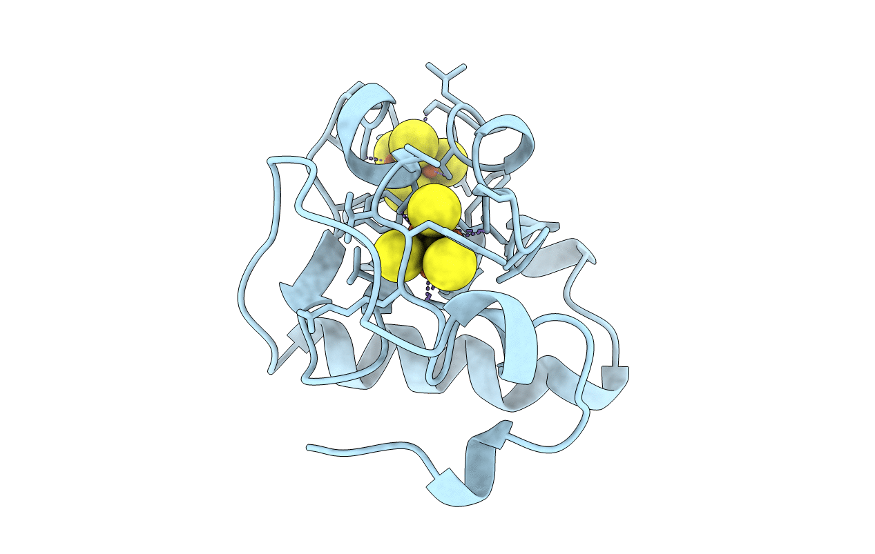
7-FE FERREDOXIN FROM AZOTOBACTER VINELANDII, NA DITHIONITE REDUCED, PH 8.5, 1.4A RESOLUTION, 100 K
Biological Source:
Source Organism:
PDB Version:
Deposition Date:
1998-12-11
Release Date:
1998-12-16
Method Details:
Experimental Method:
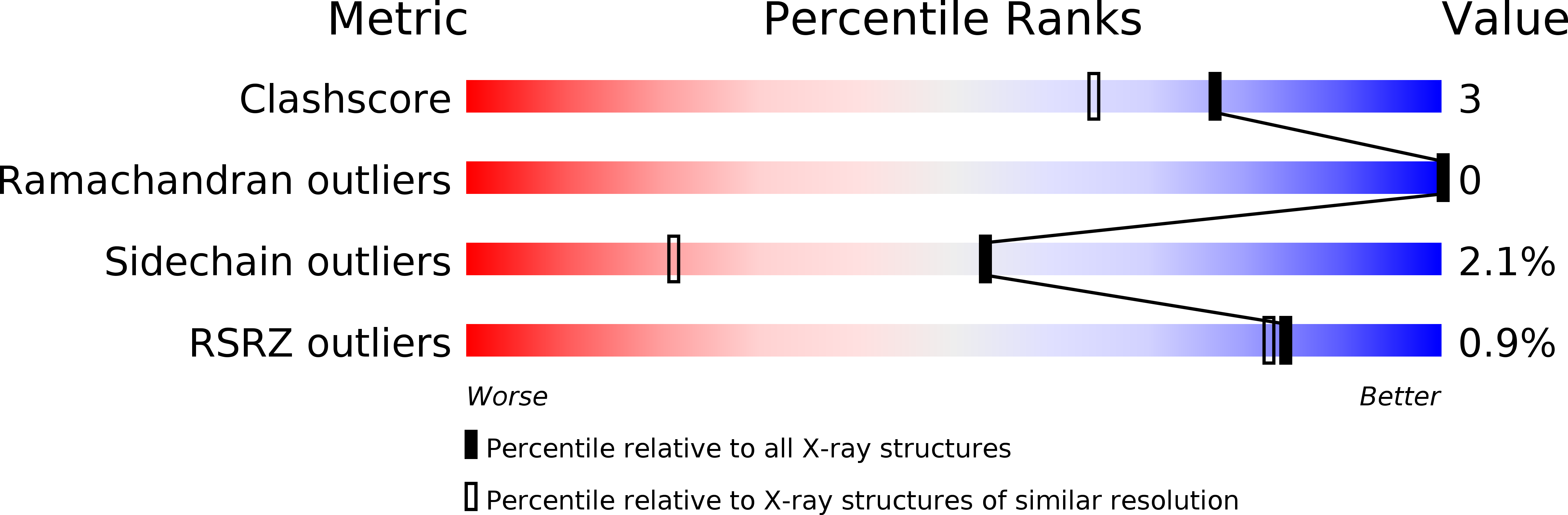
Resolution:
1.40 Å
R-Value Observed:
0.17
Space Group:
P 41 21 2
Macromolecular Entities
Polymer Type:polypeptide(L)
Description:PROTEIN (7-FE FERREDOXIN I)
Chain IDs:A
Chain Length:106
Number of Molecules:1
Biological Source:Azotobacter vinelandii
Primary Citation
Oxidized and reduced Azotobacter vinelandii ferredoxin I at 1.4 A resolution: conformational change of surface residues without significant change in the [3Fe-4S]+/0 cluster.
Biochemistry 38 8228 8239 (1999)
PMID: 10387068 DOI: 10.1021/bi983008i

Abstact

The refined structure of reduced Azotobacter vinelandii 7Fe ferredoxin FdI at 100 K and 1.4 A resolution is reported, permitting comparison of [3Fe-4S]+ and [3Fe-4S]0 clusters in the same protein at near atomic resolution. The reduced state of the [3Fe-4S]0 cluster is established by single-crystal EPR following data collection. Redundant structures are refined to establish the reproducibility and accuracy of the results for both oxidation states. The structure of the [4Fe-4S]2+ cluster in four independently determined FdI structures is the same within the range of derived standard uncertainties, providing an internal control on the experimental methods and the refinement results. The structures of the [3Fe-4S]+ and [3Fe-4S]0 clusters are also the same within experimental error, indicating that the protein may be enforcing an entatic state upon this cluster, facilitating electron-transfer reactions. The structure of the FdI [3Fe-4S]0 cluster allows direct comparison with the structure of a well-characterized [Fe3S4]0 synthetic analogue compound. The [3Fe-4S]0 cluster displays significant distortions with respect to the [Fe3S4]0 analogue, further suggesting that the observed [3Fe-4S]+/0 geometry in FdI may represent an entatic state. Comparison of oxidized and reduced FdI reveals conformational changes at the protein surface in response to reduction of the [3Fe-4S]+/0 cluster. The carboxyl group of Asp15 rotates approximately 90 degrees, Lys84, a residue hydrogen bonded to Asp15, adopts a single conformation, and additional H2O molecules become ordered. These structural changes imply a mechanism for H+ transfer to the [3Fe-4S]0 cluster in agreement with electrochemical and spectroscopic results.

Legend

Protein

Chemical

Disease

Primary Citation of related structures