Deposition Date
2021-07-16
Release Date
2021-09-08
Last Version Date
2024-10-30
Entry Detail
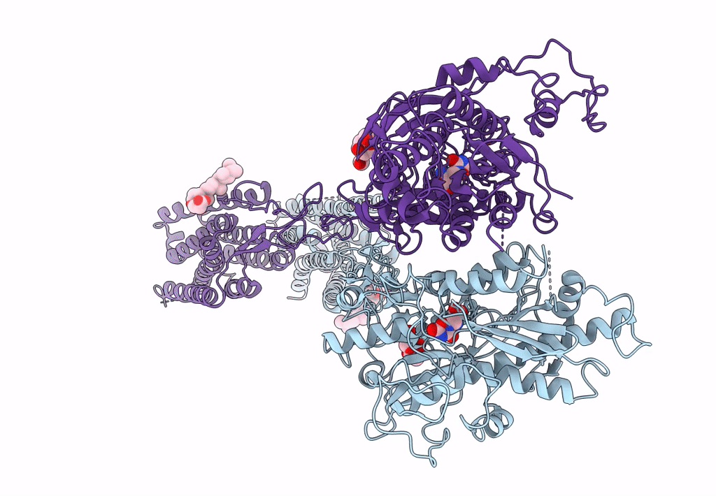
PDB ID:
7FD8
Keywords:
Title:
Thermostabilised full length human mGluR5-5M bound with L-quisqualic acid
Biological Source:
Source Organism(s):
Homo sapiens (Taxon ID: 9606)
Expression System(s):
Method Details:
Experimental Method:
Resolution:
3.80 Å
Aggregation State:
PARTICLE
Reconstruction Method:
SINGLE PARTICLE