7FCA image
Deposition Date 2021-07-14
Release Date 2021-08-04
Last Version Date 2025-03-12
Entry Detail
PDB ID:
7FCA
Keywords:
Title:
PfkB(Mycobacterium marinum)
Biological Source:
Host Organism:
Method Details:
Experimental Method:
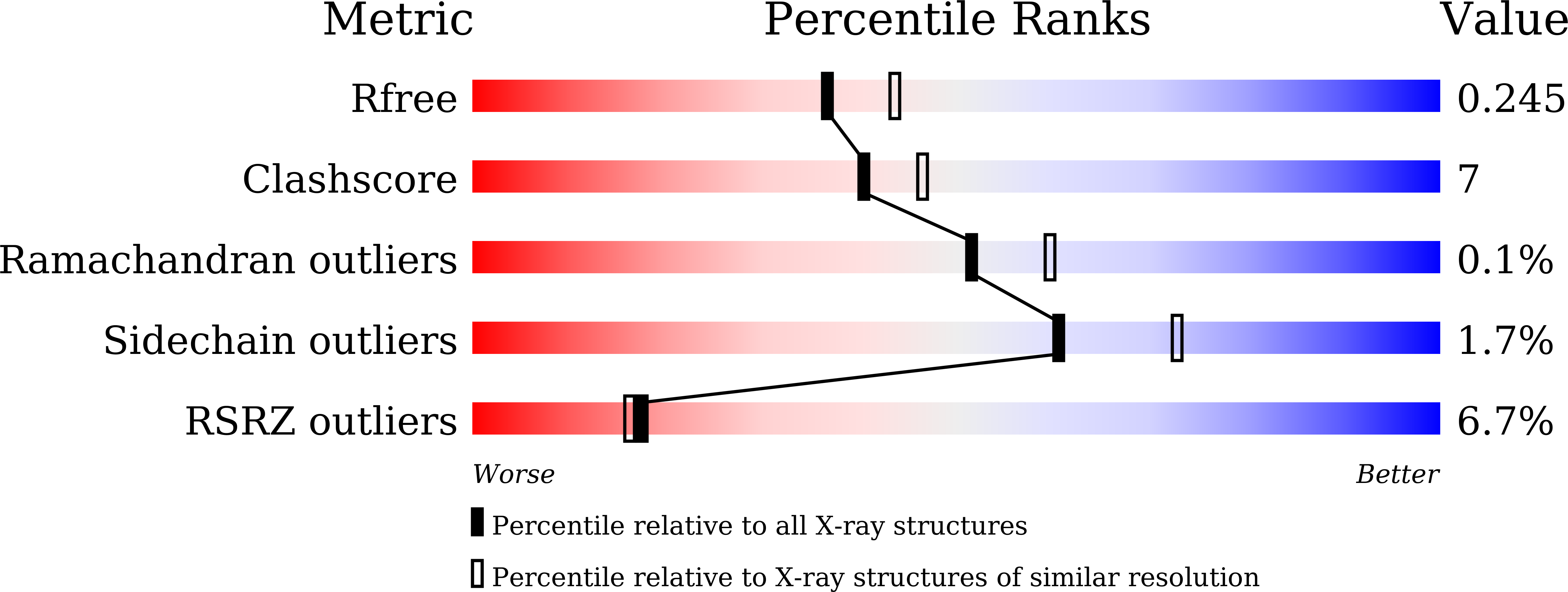
Resolution:
2.21 Å
R-Value Free:
0.24
R-Value Work:
0.21
R-Value Observed:
0.21
Space Group:
C 1 2 1
Macromolecular Entities
Polymer Type:polypeptide(L)
Molecule:Fructokinase, PfkB
Gene (Uniprot):pfkB
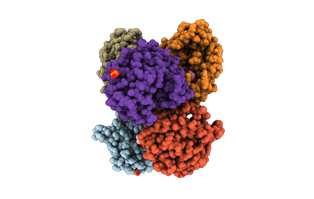
Chain IDs:A, B, C, E, F
Chain Length:303
Number of Molecules:5
Biological Source:Mycobacterium marinum (strain ATCC BAA-535 / M)
Polymer Type:polypeptide(L)
Molecule:Fructokinase, PfkB
Gene (Uniprot):pfkB
Chain IDs:D
Chain Length:291
Number of Molecules:1
Biological Source:Mycobacterium marinum (strain ATCC BAA-535 / M)
Primary Citation
Structural analysis and functional study of phosphofructokinase B (PfkB) from Mycobacterium marinum.
Biochem.Biophys.Res.Commun. 579 129 135 (2021)
PMID: 34597996 DOI: 10.1016/j.bbrc.2021.09.051

Abstact

Phosphofructokinase B (PfkB) belongs to the ribokinase family, which uses the phosphorylated sugar as substrate, and catalyzes fructose-6-phosphate into fructose-1,6-diphosphate. However, the structural basis of Mycobacterium marinum PfkB is not clear. Here, we found that the PfkB protein was monomeric in solution, which was different from most enzymes in this family. The crystal structure of PfkB protein from M. marinum was solved at a resolution of 2.21 Å. The PfkB structure consists of two domains, a major three-layered α/β/α sandwich-like domain characteristic of the ribokinase-like superfamily, and a second domain composed of four-stranded β sheets. Structural comparison analysis suggested that residues G236, A237, G238, and D239 could be critical for ATP catalysis and substrate binding of PfkB. Our current work provides new insights into understanding the mechanism of the glycolysis in M. marinum.

Legend

Protein

Chemical

Disease

Primary Citation of related structures