
Deposition Date
2021-06-28
Release Date
2022-05-11
Last Version Date
2025-05-14
Entry Detail
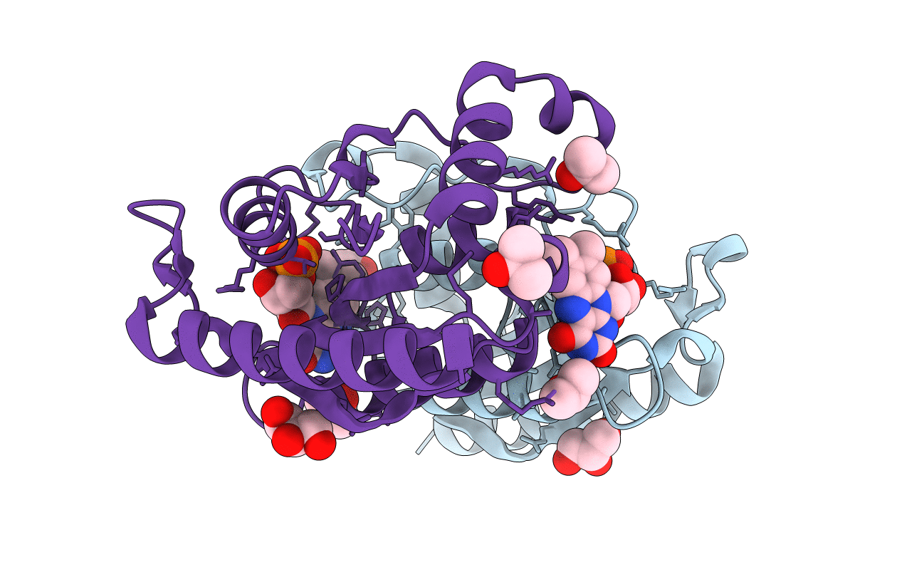
PDB ID:
7F76
Keywords:
Title:
Crystal Structure of FMN-dependent NADPH-quinone reductase (azoR) from Bacillus cohnii
Biological Source:
Source Organism(s):
Bacillus cohnii (Taxon ID: 33932)
Expression System(s):
Method Details:
Experimental Method:
Resolution:
1.57 Å
R-Value Free:
0.19
R-Value Work:
0.16
R-Value Observed:
0.16
Space Group:
P 2 21 21
