Deposition Date
2021-06-26
Release Date
2022-05-11
Last Version Date
2023-11-29
Entry Detail
PDB ID:
7F71
Keywords:
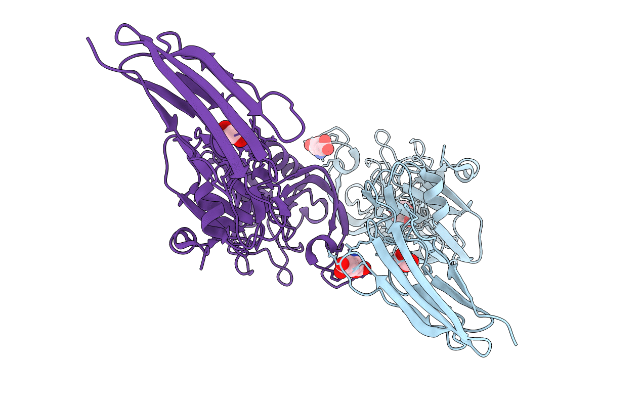
Title:
Crystal structure of the Mycobacterium tuberculosis L,D-transpeptidase-2 (LdtMt2) with peptidoglycan sugar moiety and glutamate
Biological Source:
Source Organism:
Host Organism:
Method Details:
Experimental Method:
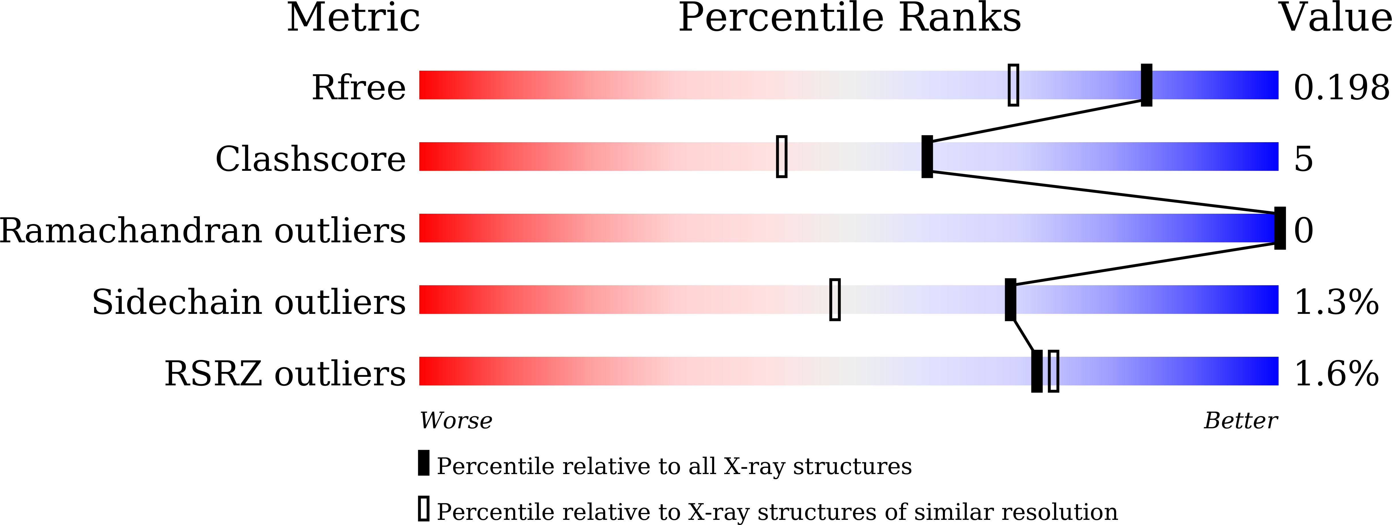
Resolution:
1.58 Å
R-Value Free:
0.19
R-Value Work:
0.16
R-Value Observed:
0.16
Space Group:
P 1 21 1